Fundus image blood vessel segmentation method based on coupled neural network and line connector

A technology of coupled neural network and line connector, which is applied in the field of fundus image blood vessel segmentation based on coupled neural network and line connector, can solve problems such as noise interference, weak contrast, uneven illumination, etc., and achieve reduced complexity, clear and complete The effect of blood vessel edges, avoiding blurring and distortion of image edges

Active Publication Date: 2020-11-06
SHANGHAI MARITIME UNIVERSITY
View PDF5 Cites 4 Cited by
  • Summary
  • Abstract
  • Description
  • Claims
  • Application Information

AI Technical Summary

Problems solved by technology

[0004] Due to the complex structure of the fundus retinal image, there are still problems such as uneven illumination, weak contrast, and noise interference, which lead to the inability of general image segmentation algorithms to segment retinal blood vessels. Finding an effective blood vessel segmentation method is a common concern of scholars today.

Method used


Image

  • Fundus image blood vessel segmentation method based on coupled neural network and line connector
  • Fundus image blood vessel segmentation method based on coupled neural network and line connector
  • Fundus image blood vessel segmentation method based on coupled neural network and line connector

Examples


Embodiment Construction

[0033] The specific implementation manner of the present invention will be described in detail below in conjunction with the accompanying drawings. The present invention can be implemented in a variety of other ways than those described here, and all other embodiments obtained by those skilled in the art without creative efforts belong to the protection scope of the present invention.

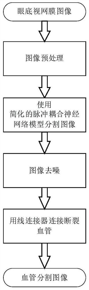
[0034] An embodiment of the present invention provides a method for segmenting blood vessels in fundus images based on a coupled neural network and a line connector. The implementation flow chart is as follows figure 1 As shown, it is mainly divided into the following steps:

[0035] 1. Obtain the fundus image to be segmented and perform preprocessing on it.

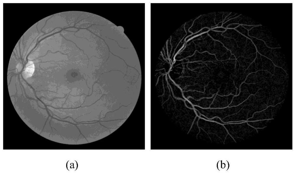
[0036] Due to the complex structure of the fundus image, there are also problems such as uneven illumination, weak contrast, and noise interference. Therefore, it is necessary to preprocess the fundus image to eliminate noise, enhance th...


PUM


Abstract

The invention discloses a fundus image blood vessel segmentation method based on a coupled neural network and a line connector and belongs to the technical field of medical image and digital image processing. The method comprises the following steps that a simplified pulse coupling neural network model is proposed, and a basic structure of a blood vessel in an image is obtained by utilizing the similarity of adjacent neurons; a new denoising method is provided, most noise points are removed through pixel connectivity, and meanwhile complete blood vessel edges are reserved, a problem of blood vessel breakage in the segmentation process is solved through the line connector so that a complete blood vessel structure is presented, and blood vessel recognition accuracy is improved. A test is carried out on two public retina databases of DRIVE and STARE, and a result shows that compared with an existing method, the method is advantaged in that excellent scores are obtained on indexes such asaverage accuracy and sensitivity, and the good response time is achieved.

Description

technical field [0001] The invention relates to the field of medical image processing, in particular to a fundus image blood vessel segmentation method based on a coupled neural network and a line connector. Background technique [0002] Changes in the structural characteristics of fundus images can reflect the existence of certain physical diseases to a certain extent. Blood vessels are the most important structure in fundus images. The diameter, color and curvature of blood vessels are closely related to the existence of diseases. Vascular and coronary artery disease and retinal lesions in infants often cause changes in the shape of retinal blood vessels. Therefore, the analysis of fundus images is crucial for the diagnosis of ophthalmic problems and other diseases such as diabetes and hypertension. Due to the time-consuming and labor-intensive manual analysis, an excellent medical image-aided diagnosis system is now needed to pre-process the image, such as automatically ...

Claims


Application Information

Patent Timeline
Patent Type & Authority Applications(China)
IPC IPC(8): G06T7/136G06T7/194G06T5/00
CPCG06T7/136G06T7/194G06T5/002G06T2207/20192G06T2207/20104G06T2207/20036G06T2207/30041G06T2207/20084
Inventor 刘锋黄林媛
Owner SHANGHAI MARITIME UNIVERSITY
Why Eureka
Try Eureka