Method and system for imaging fetal brain channeling three-dimensional image in ultrasonic image

An ultrasonic image, three-dimensional technology, applied in the direction of ultrasonic/sonic/infrasonic image/data processing, ultrasonic/sonic/infrasonic Permian technology, image enhancement, etc., can solve the problem of accurate observation of the surface of the brain sulci and clear images The problem of poor precision and accuracy, inconsistent detection results, etc., to eliminate the impact of overlapping tissue on the image, to solve the high professional level requirements, to solve the effect of low clarity and accuracy

Pending Publication Date: 2020-11-13
SHENZHEN MATERNITY & CHILD HEALTHCARE HOSPITAL
View PDF0 Cites 2 Cited by
  • Summary
  • Abstract
  • Description
  • Claims
  • Application Information

AI Technical Summary

Benefits of technology

This technology allows us to create 3D models from an ultrasound dataset containing multiple layers of grayscale values obtained during medical procedures or examinations without having to manually adjust each layer separately. It also helps improve visualization quality when viewing these 3D model structures under realistic conditions.

Problems solved by technology

The technical problem addressed by this patented method relating to neonate cardiovascular disease (NCL) is that current noninvasive techniques cannot accurately predict or distinguish between different types of neural tissue structures associated with NCL from those seen before delivery without surgery. This results in increased patient risk due to complications caused by incorrect treatment when delivering newborns.

Method used


Image

  • Method and system for imaging fetal brain channeling three-dimensional image in ultrasonic image
  • Method and system for imaging fetal brain channeling three-dimensional image in ultrasonic image
  • Method and system for imaging fetal brain channeling three-dimensional image in ultrasonic image

Examples


Embodiment Construction

[0065] In order to make the purpose, technical solution and advantages of the present invention clearer, the present invention will be further described in detail below in conjunction with the accompanying drawings and embodiments. It should be understood that the specific embodiments described here are only used to explain the present invention, not to limit the present invention. In addition, the technical features involved in the various embodiments of the present invention described below can be combined with each other as long as they do not constitute a conflict with each other.

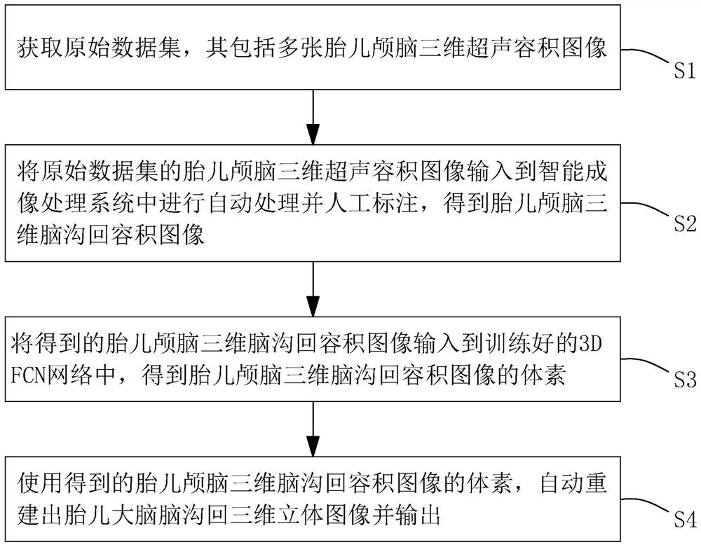
[0066] The purpose of the present invention is to provide a three-dimensional image imaging method of fetal brain sulci in ultrasound images, through deep learning a large number of normal and abnormal fetal brain three-dimensional ultrasound volume image data in early pregnancy, to realize automatic generation of fetal brain sulci 3D images to assess fetal brain development more intuitively an...


PUM


Abstract

The invention discloses a method for imaging fetal brain channeling three-dimensional image in an ultrasonic image, which aims to quickly and visually evaluate the development condition of fetal brainchanneling by adopting an artificial intelligence method through three-dimensional volume data of fetal brain. The method comprises the following steps: obtaining a large amount of three-dimensionalultrasonic volume data of fetal brain; inputting volume data to an intelligent imaging processing system, and manually marking brain channeling so as to obtain fetal craniocerebral three-dimensional brain channeling volume image data; inputting the fetal craniocerebral three-dimensional brain channeling volume image data into a trained 3D FCN network to obtain voxels of a fetal craniocerebral three-dimensional brain channeling volume image; automatically reconstructing a fetal craniocerebral channeling three-dimensional image by using voxels of the fetal craniocerebral three-dimensional channeling volume image, and outputting the fetal craniocerebral channeling three-dimensional image. The method can solve the technical problems that an existing clinician cannot reconstruct a fetal brain channeling three-dimensional image, the reconstruction steps are complex, the reconstruction individual difference is large, an objective analysis basis is lacked, and the result repeatability is poor.

Description


Claims


Application Information

Patent Timeline
Owner SHENZHEN MATERNITY & CHILD HEALTHCARE HOSPITAL
Why Eureka
Try Eureka