Liver lesion image processing method and system, storage medium, program and terminal

An image processing and liver technology, applied in the field of image processing, can solve problems such as lack of effect, and achieve the effect of improving accuracy and improving accuracy

Inactive Publication Date: 2020-11-17
十堰市太和医院(湖北医药学院附属医院)
View PDF5 Cites 6 Cited by
  • Summary
  • Abstract
  • Description
  • Claims
  • Application Information

AI Technical Summary

Problems solved by technology

Traditional methods rely on feature engineering for lesion recognition on small data sets, and the effect is not as good as that based on deep learning

Method used


Image

  • Liver lesion image processing method and system, storage medium, program and terminal
  • Liver lesion image processing method and system, storage medium, program and terminal
  • Liver lesion image processing method and system, storage medium, program and terminal

Examples


Embodiment Construction

[0053] In order to make the object, technical solution and advantages of the present invention more clear, the present invention will be further described in detail below in conjunction with the examples. It should be understood that the specific embodiments described here are only used to explain the present invention, not to limit the present invention.

[0054] Aiming at the problems existing in the prior art, the present invention provides a liver lesion image processing method, system, storage medium, program, and terminal. The present invention will be described in detail below with reference to the accompanying drawings.

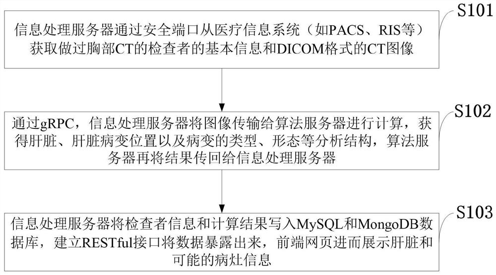
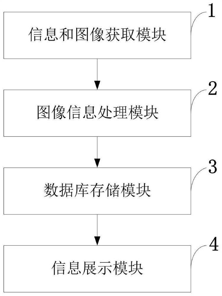
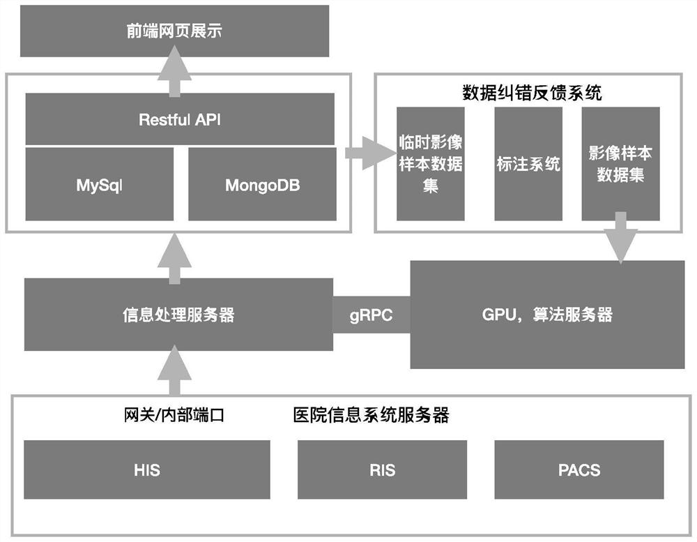
[0055] Such as figure 1 As shown, the liver lesion image processing method provided by the present invention includes the following main steps:

[0056] S101: the information processing server obtains the basic information of the examiner who has undergone chest CT and the CT image in DICOM format from the medical information system (such as PACS, RI...


PUM


Abstract

The invention belongs to the technical field of image processing, and discloses a liver lesion image processing method and system, a storage medium, a program and a terminal. An information processingserver acquires basic information and a CT image of an examiner who has undergone chest CT from a security port; through a gRPC, the information processing server transmits the image to the algorithmserver for calculation, and an algorithm server transmits a result back to the information processing server; the information processing server writes the inspector information and the calculation result into a MySQL and MongoDB database, and a RESTful standard interface is established to expose the data; and a front-end webpage displays liver and possible focus information. On the basis of the existing classic neural network unet architecture, on the basis of continuously feeding back an increased liver lesion data set, through comprehensive improvement of the model, the liver recognition accuracy can be improved to 97%, and the liver occupation recognition accuracy is improved to 98%.

Description

technical field [0001] The invention belongs to the technical field of image processing, and in particular relates to a liver lesion image processing method, system, storage medium, program, and terminal. Background technique [0002] At present, among the major industries of artificial intelligence, the direction of medical treatment plus artificial intelligence has become the focus of attention and has attracted much attention. The combination of artificial intelligence and medical treatment has extraordinary development potential and imagination. Computer vision and medical imaging have always been closely related. Driven by the rapid development of AI technology, a large number of imaging diagnostic products have emerged on the market, such as lung nodule detection, bone and joint fracture and bone age detection, cerebral hemorrhage and brain tumor detection, fundus Disease screening, cardiovascular examination, and even the recent detection of new coronary pneumonia an...

Claims


Application Information

Patent Timeline
Patent Type & Authority Applications(China)
IPC IPC(8): G06K9/62G06T7/00
CPCG06T7/0012G06T2207/10081G06T2207/20081G06T2207/30056G06F18/214
Inventor 赵亮徐霖罗杰陈文何强高瞻王克军胡瑞张涛姜鹏
Owner 十堰市太和医院(湖北医药学院附属医院)
Why Eureka
Try Eureka