Image segmentation method for kidney tumor

An image segmentation and renal tumor technology, applied in the field of image segmentation of renal tumors, can solve the problems of easy interference from other organs and tissues, low accuracy, large search space, etc., to enhance feature learning ability and improve accuracy. , the effect of narrowing the search space

Pending Publication Date: 2020-12-15
XIAMEN UNIV
View PDF0 Cites 5 Cited by
  • Summary
  • Abstract
  • Description
  • Claims
  • Application Information

AI Technical Summary

Problems solved by technology

At present, CT imaging examination is one of the main examination methods for renal tumors and other renal diseases. According to the size of renal tumors, doctors can grade the severity of tumors and formulate corresponding treatment methods; at the same time, they can locate renal tumors and analyze their shape and size; the existing accurate segmentation and judgment of the kidney and renal tumor area on the acquired kidney image through medical image processing has effectively alleviated the workload of doctors and demonstrated the effectiveness of technology intelligence. However, currently in the image segmentation task Most of them are implemented in an end-to-end manner, that is, a complete image is input at one time, the network returns the segmentation results of the whole image, and the sliding window is used to traverse the complete image in sequence, and the segmentation network performs local regions corresponding to each sliding window. Carry out segmentation prediction, and finally combine the local segmentation results of all small windows into a complete global image segmentation result according to the spatial position and traversal order. For tumor segmentation and judgment, not only the search space is large but also it is easily interfered by other organs and tissues, so the efficiency is low and the accuracy is not high

Method used


Image

  • Image segmentation method for kidney tumor
  • Image segmentation method for kidney tumor
  • Image segmentation method for kidney tumor

Examples


Embodiment

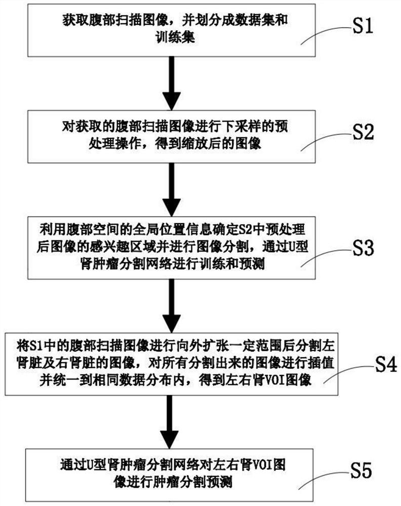
[0033] Cooperate Figure 1 to 6 As shown, the present invention discloses an image segmentation method of a kidney tumor, comprising the steps of:

[0034] S1, get the abdomen scan image, and divide it into a data set and training set.

[0035] S2, the acquired abdomen scanned image is subjected to the pretreatment operation to obtain a scaled image.

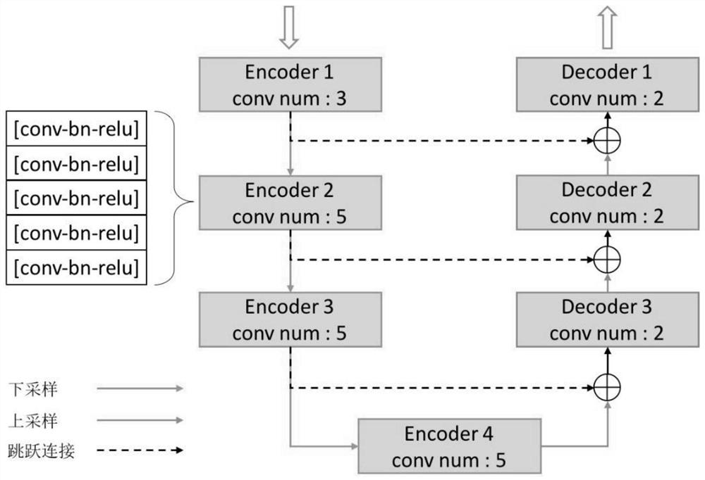
[0036] S3, using the global location information of the abdomen space to determine the region of interest in S2, and image segmentation, and perform training and prediction through the U-type renal tumor division network.

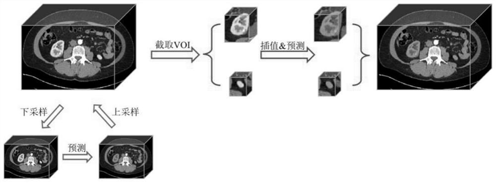
[0037] S4, the abdominal scanning image in S1 is expanded outwardly and divided the image of the left kidney and the right kidney, interpolating all divided images and uniform to the same data distribution, to obtain the left and right kidney VOI images.

[0038] S5, tumor segmentation prediction of left and right kidney VOI images by U-type renal tumor splitting network.

[0039] The common CT, MRI data is common...


PUM


Abstract

The invention discloses an image segmentation method for a kidney tumor, and the method comprises the following steps: S1, obtaining an abdomen scanning image, and dividing the abdomen scanning imageinto a data set and a training set; S2, performing down-sampling preprocessing on the acquired abdomen scanning image to obtain a scaled image; S3, determining an area of interest of the preprocessedimage in the step S2 by using global position information of the abdominal space, performing image segmentation, and performing training and prediction by a U-shaped kidney tumor segmentation network;S4, outwards expanding the abdomen scanning image in the step S1 for a certain range, segmenting images of the left kidney and the right kidney, interpolating all segmented images, and unifying the interpolated images into the same data distribution to obtain left and right kidney VOI images; S5, performing tumor segmentation prediction on the left and right kidney VOI images by a U-shaped kidneytumor segmentation network. Interference of other organs and tissues is effectively avoided, the accuracy of kidney tumor identification and image segmentation is improved, and efficiency is higher.

Description

Technical field [0001] The present invention relates to the technical field of medical image processing, and more particularly to an image segmentation method of a renal tumor. Background technique [0002] The kidney is an important organ of the human body. Once the renal function is damaged, it will lead to a variety of metabolic final products to accumulate in the body, which in turn affects life safety. In various kidney diseases, the renal tumor is a top dangerous disease of kidney health. At present, CT imaging examination is one of the main inspection methods of kidney disease such as renal tumors. According to the size of the renal tumor, the doctor can grade the severity of the tumor, and formulate corresponding treatment; at the same time, the renal tumor is positioned, and the shape is analyzed And size; existing medical image processing for accurate segmentation judgment of kidney and renal tumor regions through medical image processing, effectively alleviating the wo...

Claims


Application Information

Patent Timeline
Patent Type & Authority Applications(China)
IPC IPC(8): G06T7/11G06T9/00G06N3/04G06N3/08
CPCG06T7/11G06T9/002G06N3/08G06T2207/20081G06T2207/20084G06T2207/30084G06T2207/30096G06T2207/10081G06T2207/10088G06N3/045
Inventor 王连生
Owner XIAMEN UNIV
Why Eureka
Try Eureka