
Resolution enhancement method for ultrasonic microscope

A resolution enhancement, ultrasonic microscope technology, applied in the analysis of solids using sonic/ultrasonic/infrasonic waves, material analysis using sonic/ultrasonic/infrasonic waves, and analysis of materials, etc., can solve the problem of difficulty in improving imaging resolution.

Pending Publication Date: 2020-12-22
UNIVERSITY OF CHINESE ACADEMY OF SCIENCES
View PDF10 Cites 0 Cited by
  • Summary
  • Abstract
  • Description
  • Claims
  • Application Information

AI Technical Summary

Problems solved by technology

[0003] For this reason, the present invention provides a resolution enhancement method for ultrasonic microscopes to solve the problem that the imaging resolution of existing ultrasonic microscopes is difficult to improve

Method used


Image

  • Resolution enhancement method for ultrasonic microscope
  • Resolution enhancement method for ultrasonic microscope
  • Resolution enhancement method for ultrasonic microscope

Examples


Embodiment

[0039] This embodiment discloses a method for enhancing the resolution of an ultrasonic microscope, the method is:

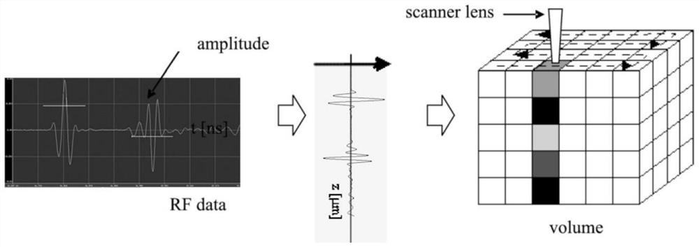
[0040] Step 1, setting parameters to obtain the ultrasonic tomographic scanning results of the test piece;

[0041] Step 2, adopt discrete wavelet decomposition to carry out multi-scale analysis to the ultrasonic signal of each point, generate low-frequency component and high-frequency component;

[0042] Step 3, use the Wiener deconvolution filter to process the low frequency components, and separate the superimposed signals;

[0043] Step 4, performing discrete wavelet inverse transform on the separated signal and high-frequency component, and reconstructing the ultrasonic tomographic scanning data;

[0044] Step 5. Using the reconstructed ultrasound tomography data to generate higher resolution vertical and horizontal profile images.

[0045] In step 1, set the parameters to obtain the ultrasonic tomographic scanning data of the specimen, and its data struc...


PUM


Abstract

The invention discloses a resolution enhancement method for an ultrasonic microscope. The method comprises the following steps: firstly, acquiring ultrasonic tomography data of a test piece; carryingout multi-scale analysis on the ultrasonic signal of each point by adopting discrete wavelet decomposition to generate a low-frequency component and a high-frequency component; processing the low-frequency components by using a Wiener deconvolution filter, and separating the superposed signals; conducting discrete wavelet inverse transformation by using the separated signals and high-frequency components to reconstruct ultrasonic tomography data; and generating vertical and horizontal profile scan images of higher resolution in the test piece by using the reconstructed ultrasound tomography scan data. The problem that the imaging resolution of an existing ultrasonic microscope is difficult to improve is solved.

Description

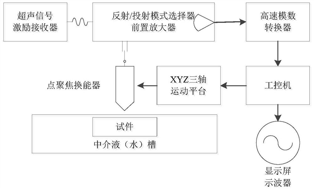
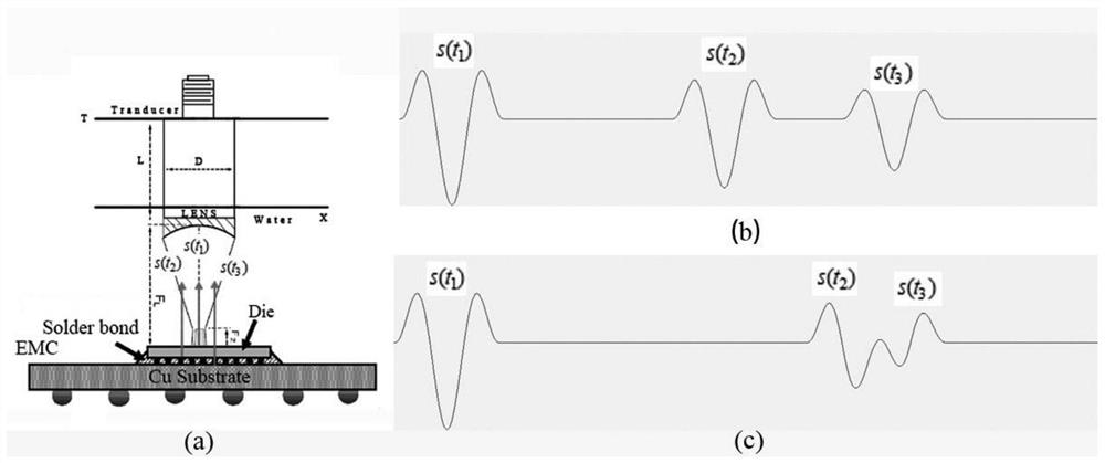
technical field [0001] The invention relates to the fields of acoustic technology and nondestructive testing technology, in particular to a resolution enhancement method for ultrasonic microscopes. Background technique [0002] Ultrasonic microscopes are non-destructive testing and microscopic imaging equipment widely used in high-tech manufacturing industries such as chip manufacturing, biomedicine, material science, and aerospace. Ultrasound microscope (if attached figure 1 ) generally use liquid-immersion point-focused ultrasonic transducers (as attached figure 2 ), the transducer can converge the sound beam at one point, so that the energy in the convergence area is concentrated and strengthened, and the width of the sound beam becomes smaller, which can meet the detection requirements of high sensitivity and high resolution. With the continuous improvement of the chip manufacturing process, the layer thickness inside the chip has been lower than the wavelength of the...

Claims


Application Information

Patent Timeline
IPC IPC(8): G01N29/06G01N29/44G06F17/14
CPCG01N29/0681G01N29/0672G01N29/44G01N29/4472G06F17/148G06F17/141
Inventor 梁昊吕科薛健
Owner UNIVERSITY OF CHINESE ACADEMY OF SCIENCES
Learn More