
Image correction method and device and capsule endoscope

A capsule endoscope and image correction technology, applied in the field of medical devices, can solve problems affecting the accuracy of capsule endoscope detection results, affecting image recognition, unfavorable focus identification and diagnosis, etc.

Active Publication Date: 2022-07-08
SHENZHEN JIFU MEDICAL TECH CO LTD
View PDF11 Cites 0 Cited by
  • Summary
  • Abstract
  • Description
  • Claims
  • Application Information

AI Technical Summary

Problems solved by technology

[0002] When the capsule endoscope collects images from the digestive tract of the subject, it will be affected by the magnetic force outside the body and the liquid in the body. While satisfying the image quality, it will cause a certain amount of smear under different exposure delay times. The current solution to smear The method of shadowing is to sharpen and enhance the edge of the image in the spatial domain. This method will be affected by the range of the operating neighborhood and the gradient operator, and will be caused by problems such as the neighborhood is too small or too large, and the degree of operator fitting. A certain degree of edge distortion cannot remove the blurred jitter based on the actual movement of the capsule endoscope, thus affecting image recognition, which is not conducive to the identification and diagnosis of lesions, and thus affects the accuracy of the detection results of the capsule endoscope

Method used


Image

  • Image correction method and device and capsule endoscope
  • Image correction method and device and capsule endoscope
  • Image correction method and device and capsule endoscope

Examples


Embodiment Construction

[0065] In order to make the objectives, technical solutions and advantages of the present invention clearer, the present invention will be further described in detail below with reference to the accompanying drawings and embodiments. It should be understood that the specific embodiments described herein are only used to explain the present invention, but not to limit the present invention.

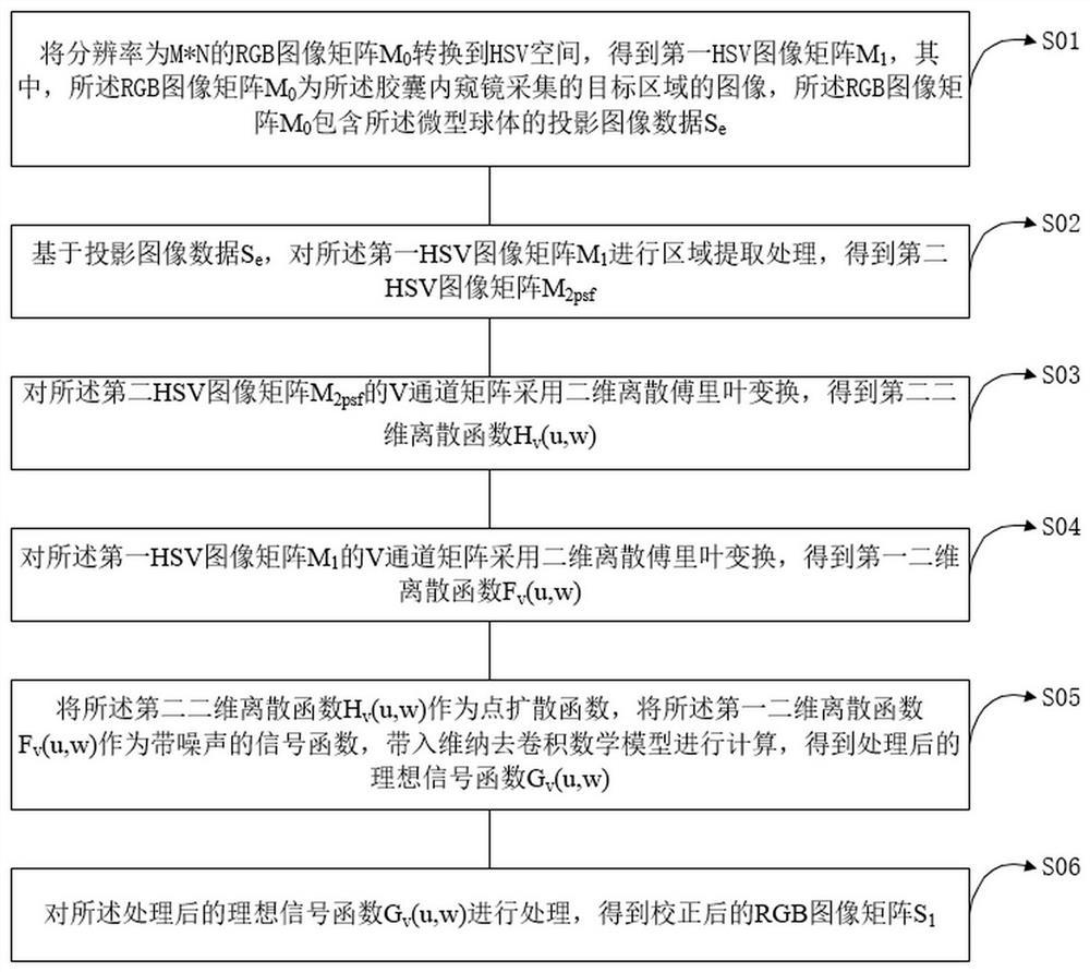
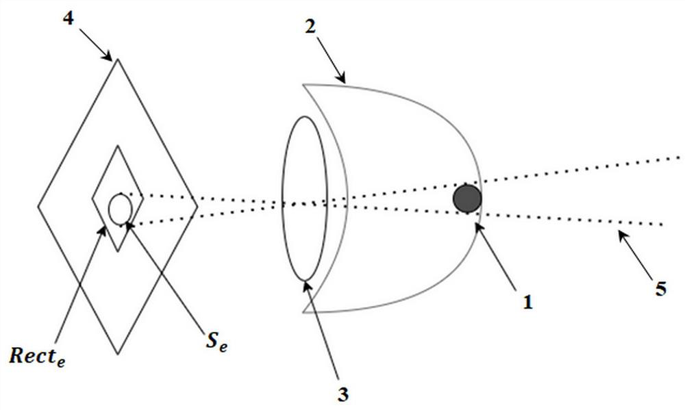
[0066] like figure 1 and figure 2 shown, figure 1 A flowchart of an image correction method provided by an embodiment of the present invention, figure 2 A schematic diagram of lighting in an image correction method provided by an embodiment of the present invention. An image correction method provided by an embodiment of the present invention is applied to a capsule endoscope. The transparent cover 2 of the capsule endoscope is provided with a microsphere 1, and the diameter d of the microsphere 1 is greater than 0.1 μm and less than 100 μm. The surface of the microsphere 1 is coated...


PUM


Abstract

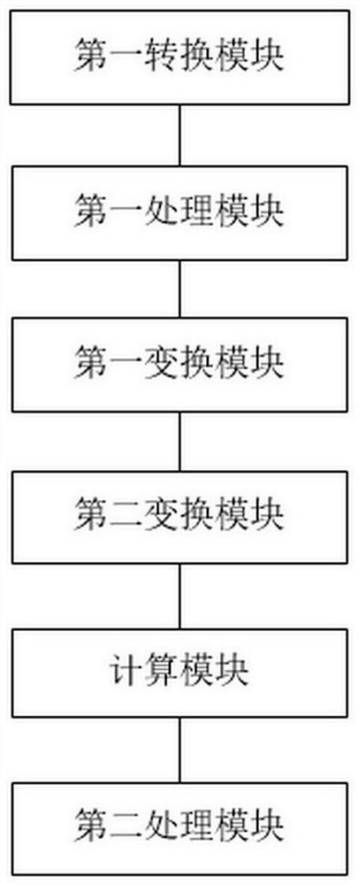
The invention discloses an image correction method and device and a capsule endoscope, and the method comprises the steps: converting an RGB image matrix M0 into an HSV space, and obtaining a first HSV image matrix M1, the M0 being an image of a target region collected by the capsule endoscope, and the M0 comprising projection image data Se of a microsphere; based on the Se, carrying out region extraction processing on the M1 to obtain a second HSV image matrix M2psf; respectively carrying out Fourier transform on the M2psf and the V channel matrix of the M1 to obtain a second two-dimensional discrete function Hv (u, w) and a first two-dimensional discrete function Fv (u, w); taking the Hv (u, w) as a point spread function, taking the Fv (u, w) as a noisy signal function, and substituting the Hv (u, w) and the Fv (u, w) into a Wiener deconvolution mathematical model for calculation to obtain a processed ideal signal function Gv (u, w); and processing the Gv (u, w) to obtain a corrected RGB image matrix S1. The fuzzy jitter is removed based on the actual motion, so that the authenticity of the image is kept, the identification and diagnosis of the focus are facilitated, and the accuracy of the detection result of the capsule endoscope is further ensured.

Description

technical field [0001] The invention relates to the technical field of medical devices, and in particular, to an image correction method, a device and a capsule endoscope. Background technique [0002] Capsule endoscope will be affected by external magnetic force and internal liquid when collecting images in the digestive tract of the subject. While satisfying the image quality, different exposure delay times will cause a certain smear. The method of shadowing is to sharpen and enhance the edge of the image in the spatial domain. This method will be affected by the range of the operation neighborhood and the gradient operator, which may be caused by problems such as the neighborhood being too small or too large and the degree of operator fitting. A certain degree of edge distortion cannot remove the blurred jitter based on the actual motion of the capsule endoscope, which affects the recognition of images, is not conducive to the identification and diagnosis of lesions, and ...

Claims


Application Information

Patent Timeline
Patent Type & Authority Applications(China)
IPC IPC(8): G06T5/00G06T5/10G06T5/40G06T7/00G06T7/90A61B1/04A61B1/00
CPCG06T5/10G06T5/40G06T7/0012G06T7/90A61B1/041A61B1/00009G06T2207/10068G06T2207/20056G06T2207/20201G06T5/80G06T5/73
Inventor 夏波阚述贤王建平
Owner SHENZHEN JIFU MEDICAL TECH CO LTD
Learn More