
Intelligent diagnosis method for rectal cancer lymph node metastasis

A lymph node metastasis and intelligent diagnosis technology, applied in neural learning methods, 2D image generation, image data processing, etc., can solve problems such as inaccurate radiomics features, low tumor segmentation accuracy, and affecting accuracy

Pending Publication Date: 2020-12-25
YANCHENG INST OF TECH
View PDF0 Cites 10 Cited by
  • Summary
  • Abstract
  • Description
  • Claims
  • Application Information

AI Technical Summary

Problems solved by technology

Traditional methods of medical image segmentation mostly rely on manual segmentation by doctors. It takes at least 10 minutes to segment a patient’s CT image. Long-term reading is prone to visual fatigue, and the results are often highly subjective and misdiagnosed. Tumor segmentation accuracy Not high, resulting in inaccurate extracted radiomics features, which ultimately affects the accuracy of prediction

Method used


Image

  • Intelligent diagnosis method for rectal cancer lymph node metastasis
  • Intelligent diagnosis method for rectal cancer lymph node metastasis
  • Intelligent diagnosis method for rectal cancer lymph node metastasis

Examples


Embodiment 1

[0061] An intelligent diagnosis method and device for lymph node metastasis of rectal cancer according to an embodiment of the present invention, specifically comprising the following steps:

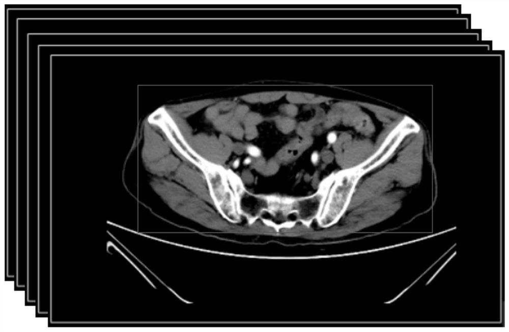
[0062] Step 1, for example figure 2 The preprocessing of the CT image data of the patient’s abdomen is shown, using the SimpleITK toolkit to read the DCM image file, using the Numpy toolkit to convert the file into a three-dimensional array matrix, using the linear normalization method to process the three-dimensional matrix data, and setting the threshold to use image intensity analysis The method removes fat and bone tissue in CT images, uses the image connected area method to eliminate the equipment background interference information in the image, uses Python slice technology to intercept the effective area in the image, and constructs the preprocessed protocol database.


PUM


Abstract

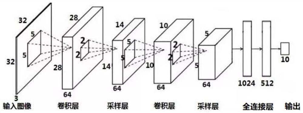
The invention discloses an intelligent diagnosis method for rectal cancer lymph node metastasis, and relates to the field of intelligent medical imaging diagnosis, and the method specifically comprises the following steps: carrying out the preprocessing of CT image data of the abdomen of a patient, reading a DCM image file, converting the file into a three-dimensional array matrix, and carrying out the data stipulation of the matrix; sending the stipulated data as a training sample to an established convolutional neural network model (CNN) for supervised learning, classifying the CT images byusing the classification model, and detecting tumor pictures contained in the images; constructing an improved AGs-Unet network model, sending the tumor original image and the tumor mask image data into the segmentation model for training, and using the model to segment a tumor area; extracting radiology characteristic data from the tumor image area, wherein the radiology characteristic data comprises texture characteristics, gray scale characteristics and morphological characteristics; selecting effective feature data to train a support vector machine SVM classification model, and using the classification model to predict and diagnose whether lymph node metastasis exists in rectal cancer or not. The rectal cancer tumor segmentation precision and lymph node metastasis diagnosis accuracy are improved.

Description

technical field [0001] The invention relates to the field of intelligent medical image diagnosis, in particular to an intelligent diagnosis method for rectal cancer lymph node metastasis. Background technique [0002] Rectal cancer refers to malignant tumors from the dentate line to the rectosigmoid junction, and is one of the most common malignant tumors of the digestive tract. In recent years in China, the incidence of rectal cancer has been increasing. Rectal cancer tends to infiltrate outside the intestine and develop lymph nodes and distant organs. [0003] Once metastases occur, patients often need to receive adjuvant chemotherapy and radiotherapy first to obtain the opportunity for surgery, and the prognosis of patients is relatively early. [0004] Patients with bowel cancer have a poor prognosis. Lymph node metastasis is one of the important factors in preoperative TNM staging. Therefore, the study of lymph node metastasis is very important for clinical medical r...

Claims


Application Information

Patent Timeline
IPC IPC(8): G06T11/00G06K9/34G06K9/62G06N3/04G06N3/08
CPCG06T11/003G06N3/084G06T2211/424G06V10/267G06N3/045G06F18/2411G06F18/24155
Inventor 王东洋
Owner YANCHENG INST OF TECH
Learn More