Medical image segmentation method and device based on multi-modal subspace clustering

A medical image and clustering method technology, which is applied in image analysis, neural learning methods, image enhancement, etc., can solve the problems of good segmentation effect, high precision, and low segmentation accuracy of complex medical images

Pending Publication Date: 2021-01-01
SOUTHWEAT UNIV OF SCI & TECH
View PDF1 Cites 12 Cited by
  • Summary
  • Abstract
  • Description
  • Claims
  • Application Information

AI Technical Summary

Problems solved by technology

In addition, many image segmentation methods based on clustering methods also have problems such as low segmentation accuracy.
[0006] The purpose of the present invention is to provide a medical image segmentation method and device based on multimodal subspace clustering, which can solve the above-mentioned problems in the complex medical image segmentation, and the complex medical image segmentation effect is good and the precision is high

Method used


Image

  • Medical image segmentation method and device based on multi-modal subspace clustering
  • Medical image segmentation method and device based on multi-modal subspace clustering
  • Medical image segmentation method and device based on multi-modal subspace clustering

Examples


Embodiment 1

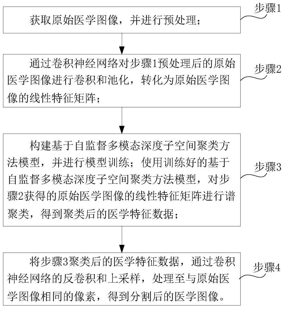
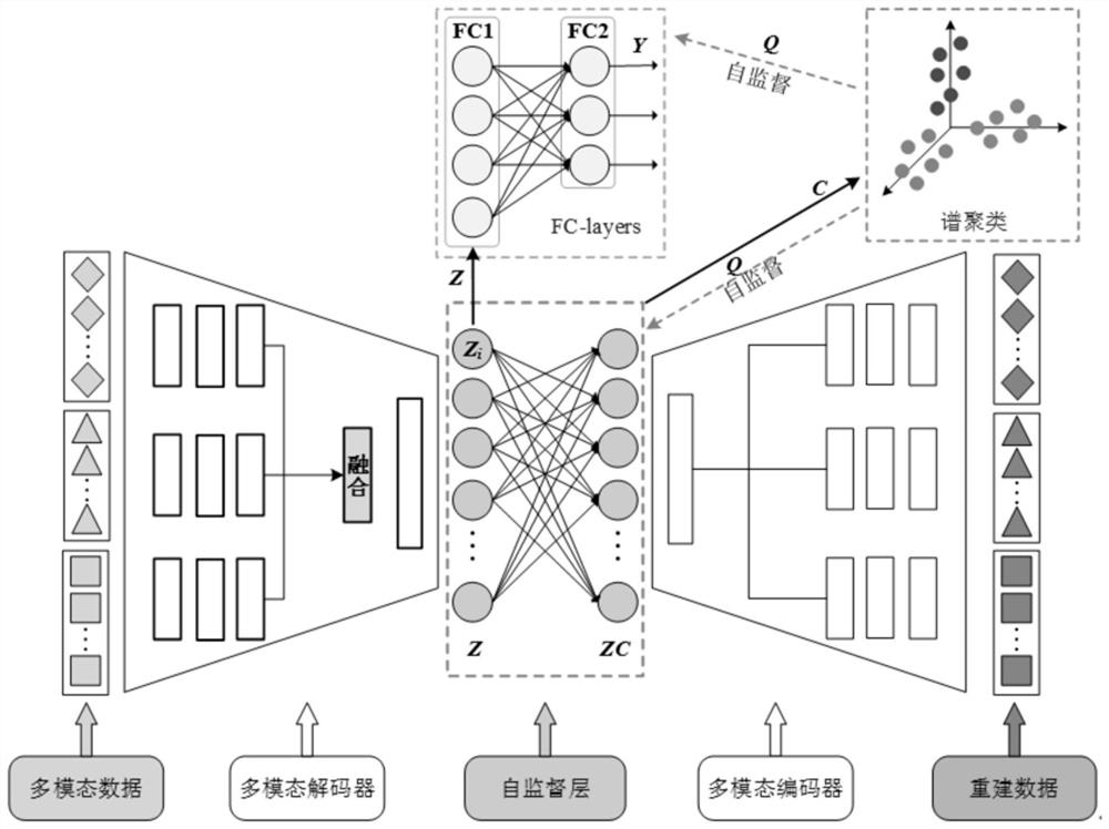
[0084] Such as Figure 1 to Figure 10 As shown, the present invention is based on the medical image segmentation method of self-supervised multimodal deep subspace clustering, the segmentation method framework is as follows figure 2 As shown, the present invention builds a model based on self-supervised multimodal deep subspace clustering method, uses useful self-supervised information in subspace clustering results to guide feature learning, and refines the self-expression model. Inspired by the deep multimodal subspace clustering algorithm, on the basis of the automatic encoding network, the feature fusion is performed after the feature extraction of the data of each modality; and then the self-expression coefficient is learned through the introduction of the self-expression layer; In particular, we will introduce a dual self-supervision that integrates feature extraction for multimodal encoding modules, affinity learning for self-representation models, and data segmentatio...

Embodiment 2

[0109] Such as Figure 1 to Figure 10 As shown, the difference between this embodiment and Embodiment 1 is that this embodiment provides a medical image segmentation device, including:

[0110] Input module: used to input the acquired original medical image;

[0111] Preprocessing module: used to preprocess the original medical image input by the input unit and send it to the processing unit;

[0112] Processing module: including a first processing unit, a second processing unit and a third processing unit;

[0113] The first processing unit is configured to perform convolution and pooling on the original medical image preprocessed by the preprocessing module through a convolutional neural network, and convert it into a linear feature matrix of the original medical image;

[0114] The second processing unit is used to construct a model based on the self-supervised multimodal deep subspace clustering method, and perform model training; using the trained self-supervised multim...

Embodiment 3

[0119] Such as Figure 1 to Figure 10 As shown, the difference between this embodiment and Embodiment 1 is that this embodiment provides a medical image segmentation device, including:

[0120] memory for storing computer programs;

[0121] A processor, configured to implement the steps of the medical image segmentation method described in Embodiment 1 when executing the computer program.


PUM


Abstract

The invention discloses a medical image segmentation method and device based on multi-modal subspace clustering, and the method comprises the steps: 1, obtaining an original medical image, and carrying out the preprocessing; 2, performing convolution and pooling on the original medical image preprocessed in the step 1 through a convolutional neural network, and converting the original medical image into a linear feature matrix of the original medical image; 3, constructing a model based on a self-supervision multi-modal depth subspace clustering method, and carrying out model training; performing spectral clustering on the linear feature matrix of the original medical image obtained in the step 2 by using a trained self-supervised multi-modal depth subspace clustering method model to obtain clustered medical feature data; and 4, processing the medical feature data clustered in the step 3 to pixels the same as those of the original medical image through deconvolution and up-sampling ofa convolutional neural network to obtain a segmented medical image. The method is good in complex medical image segmentation effect and high in precision.

Description

technical field [0001] The invention relates to the technical field of medical image segmentation, in particular to a medical image segmentation method and device based on multimodal subspace clustering. Background technique [0002] Medical magnetic resonance (MR) images are widely used in clinical medical diagnosis and research due to their advantages of high contrast, high resolution, and multi-directional. In order to effectively extract key information in images, image segmentation has become an essential link in medical image processing. However, medical image data often have high dimensions and heterogeneous features of various attributes (modalities), among which high-dimensional data generally contain more redundant features, using the existing threshold image segmentation method, edge detection image Traditional image segmentation methods such as segmentation method and regional image segmentation are not only time-consuming but often difficult to achieve good seg...

Claims


Application Information

Patent Timeline
Patent Type & Authority Applications(China)
IPC IPC(8): G06T7/10G06K9/62G06N3/04G06N3/08
CPCG06T7/10G06N3/08G06T2207/10088G06T2207/20081G06T2207/20084G06T2207/30004G06N3/045G06F18/23
Inventor 张小乾万黎明刘知贵郭丽白克强秦明伟罗亮李理
Owner SOUTHWEAT UNIV OF SCI & TECH
Why Eureka
Try Eureka