Microsurgery auxiliary device

An auxiliary device, a technology for microsurgery, applied in the direction of operating microscopes, etc., can solve the problems of high manufacturing cost, difficult to meet the requirements of low latency in microsurgery, complex structure, etc., to reduce visual fatigue, no loss of brightness, and fixed structure. simple effect

Pending Publication Date: 2021-01-15
ZUMAX MEDICAL
View PDF2 Cites 1 Cited by
  • Summary
  • Abstract
  • Description
  • Claims
  • Application Information

AI Technical Summary

Problems solved by technology

This solution also requires complex data processing of the collected images, which is difficult to meet the low-latency requirements of microsurgery. In addition, an imaging system with eight optical paths is required, which has a complex structure and high manufacturing costs.

Method used


Image

  • Microsurgery auxiliary device
  • Microsurgery auxiliary device
  • Microsurgery auxiliary device

Examples


Embodiment

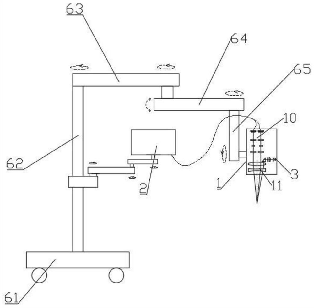
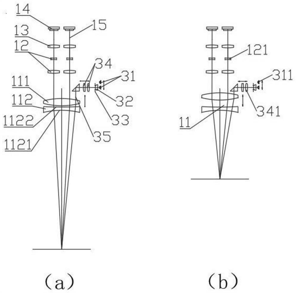
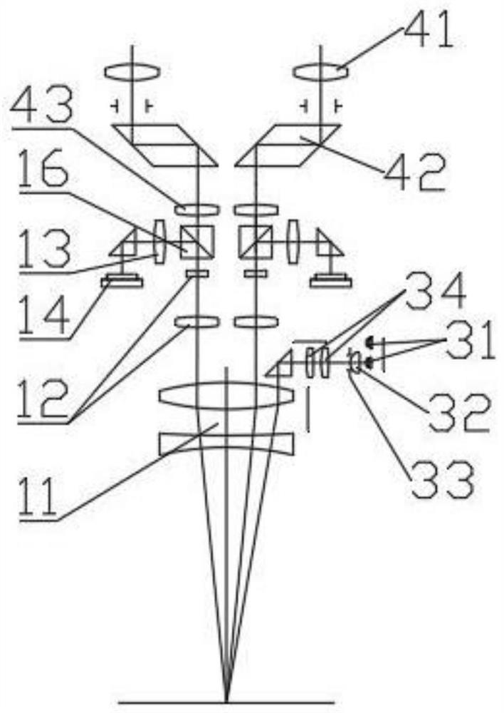
[0045]The application provides a microsurgery auxiliary device, which includes a mirror body 1 and a naked-eye 3D display 2. The lens body 1 is provided with an imaging unit 10, the imaging unit 10 includes a large objective lens group 11, a variable power lens group 12, a first barrel objective lens 13, and a photosensitive element 14. The large objective lens group 11, a variable power lens The group 12, the first barrel objective lens 13 and the photosensitive element 14 are in the same observation optical path 15 in sequence, such asfigure 1 orfigure 2 a andfigure 2 Shown in b.

[0046]The large objective lens group 11 includes at least one positive lens group 111 and at least one negative lens group 112. The positive lens group 111 and the negative lens group 112 are arranged on the same optical axis. There is a gap between the positive lens group 111 and the negative lens group 112. The distance is adjustable, and the adjustment range of the distance between the positive lens gro...


PUM


Abstract

The invention discloses a microsurgery auxiliary device. The microsurgery auxiliary device comprises a lens body and a glasses-free 3D displayer, wherein an imaging unit is arranged in the lens body,and comprises a large objective lens set, zoom lens sets, first lens cone objective lenses and photosensitive elements, and the large objective lens set, the zoom lens sets, the first lens cone objective lenses and the photosensitive elements are sequentially located on the same observation light path. The large objective lens set comprises at least one positive lens set and at least one negativelens set, the positive lens sets and the negative lens sets share the same optical axis, the distance between the positive lens sets and the negative lens sets is adjustable, the glasses-free 3D displayer is connected with the photosensitive elements, the distance between the glasses-free 3D displayer and an observer is 400-1,200 mm, and the range of the visual angle of the glasses-free 3D displayer is not less than 120 degrees. According to the microsurgery auxiliary device, the observer can directly conduct surgery operation by observing the glasses-free 3D displayer, the device is simple inoverall structure and small in system delay, the fixing mode of the glasses-free 3D displayer can be selected according to field requirements, the fixing structure is simple and reliable, an observation component can be additionally arranged when necessary, and traditional visual observation is achieved.

Description

Technical field[0001]The invention relates to the technical field of medical equipment, in particular to a microsurgery auxiliary device.Background technique[0002]Microsurgery is a delicate operation performed with the help of magnifying equipment. When the operation is performed under a traditional optical operating microscope, the tissue is magnified, not only can see the small tissues that are invisible to the naked eye during the operation, but also has a three-dimensional effect, which is beneficial to the surgeon Accurately dissect, cut and suture various tissues. However, even surgeons who are experienced in suturing blood vessels with the naked eye, without special training, are still very unaccustomed to microsurgery at the beginning of the operation. Hand-eye coordination often occurs, which affects the operation under the microscope. Therefore, it is necessary to It takes a period of training and adaptation process to skillfully perform operations under the operating micr...

Claims


Application Information

Patent Timeline
Patent Type & Authority Applications(China)
IPC IPC(8): A61B90/20A61B90/25
CPCA61B90/20A61B90/25A61B2090/309A61B2017/00216A61B2090/372
Inventor 王吉龙何进李剑月杜雷朱诚杰
Owner ZUMAX MEDICAL
Why Eureka
Try Eureka