CT image lung vessel segmentation method

A CT imaging and blood vessel technology, applied in image analysis, image enhancement, image data processing, etc., can solve the problems of generalization, poor robustness, time-consuming, high threshold requirements, etc., to ensure generalization and robustness. awesome effect

Active Publication Date: 2021-01-22
FUZHOU UNIV
View PDF9 Cites 7 Cited by
  • Summary
  • Abstract
  • Description
  • Claims
  • Application Information

AI Technical Summary

Problems solved by technology

[0004] As mentioned above, the currently commonly used pulmonary vessel segmentation method based on blood vessel enhancement requires a given threshold to distinguish pulmonary vessels from background features. The selection of the threshold has high requirements, and improper selection is prone to missing segmentation or over-segmentation; based on edge and region The deformable model needs to design the objective function of the task, and obtain the minimum energy contour under the action of internal and external forces. In complex cases, the amou

Method used


Image

  • CT image lung vessel segmentation method
  • CT image lung vessel segmentation method
  • CT image lung vessel segmentation method

Examples


Embodiment Construction

[0035] The technical solution of the present invention will be specifically described below in conjunction with the accompanying drawings.

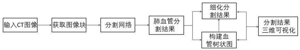
[0036] as attached figure 1 , is a flow chart of the inventive method, comprising:

[0037] 1) input image;

[0038] 2) Image preprocessing;

[0039] 3) local input image block data sampling;

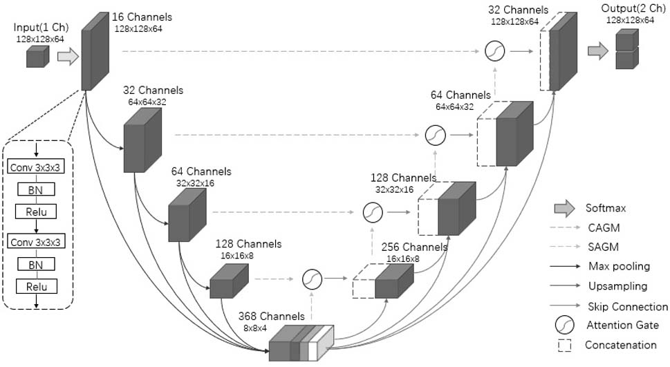
[0040] 4) Pulmonary vessel semantic segmentation network;

[0041] 5) Pulmonary vessel segmentation result map;

[0042] 6) Post-processing of the segmentation result map;

[0043] 7) Three-dimensional visualization of segmentation results;

[0044] Based on the above content, the specific implementation process is described in detail below:

[0045] First, the input image step. The invention can process CT data of different image protocols, enhanced and non-enhanced CT data.

[0046] After acquiring the image, first perform the image preprocessing step: adjust the Hounsfield unit value of all data to the range [-1024,600]HU, which contain...


PUM


Abstract

The invention relates to a CT image lung vessel segmentation method. The method comprises the steps of S1, acquiring a to-be-segmented image; s2, performing data preprocessing on the to-be-segmented image; s3, sampling a local input image block; s4, inputting the image blocks into a pre-trained segmentation model; s5, post-processing a segmentation result, and constructing a blood vessel tree diagram for refinement; and S6, carrying out three-dimensional visualization on a segmentation result. A segmentation network model is an improved 3D Unet model, and a multi-scale strategy is added to theimproved 3D Unet model to capture rich context information so as to improve the characteristic performance of pulmonary vessels. An attention mechanism is introduced for pulmonary vessel segmentationof pathological lung data, a related region of a target can be highlighted adaptively, and irrelevant features, especially the influence of a lesion region on vessel segmentation, can be inhibited implicitly.

Description

technical field [0001] The invention belongs to the fields of computer vision and digital image processing, and in particular relates to a method for segmenting pulmonary vessels in CT images. Background technique [0002] Pulmonary vessel segmentation is indispensable in computer-aided detection systems. Accurate pulmonary vessel segmentation plays an important role in clinical diagnosis, analysis of lesion and surgical planning. Using imaging, digital image processing and computer vision technology in the computer-aided detection system, the pulmonary vascular structure in the lung CT image is automatically segmented and visualized in 3D, so as to obtain the characteristic information of the target tissue structure, which can provide doctors with more information. The observation method gives doctors more references and helps doctors make faster and more accurate judgments. [0003] Traditional pulmonary vessel segmentation methods mostly rely on handcrafted features, re...

Claims


Application Information

Patent Timeline
IPC IPC(8): G06T7/10G06N3/04
CPCG06T7/10G06T2207/10081G06T2207/10012G06T2207/30101G06N3/045
Inventor 郭金泉佘宇航何炳蔚
Owner FUZHOU UNIV
Why Eureka
Try Eureka