
Patient-level tumor intelligent diagnosis method based on full-view digital slicing

A technology of digital slicing and diagnostic methods, which is applied in the field of patient-level tumor intelligent diagnosis, can solve problems such as misdiagnosis and rapid diagnosis at the difficult case level, and achieve the effects of increasing the receptive field, reducing manual operations, and enhancing capabilities

Active Publication Date: 2021-01-22
HEBEI UNIV OF TECH
View PDF10 Cites 5 Cited by
  • Summary
  • Abstract
  • Description
  • Claims
  • Application Information

AI Technical Summary

Problems solved by technology

It is easy to make a misdiagnosis of "case health" for this part of the slide when only targeting tissue tiles or full-field digital slides
In summary, existing technologies are difficult to perform rapid diagnosis at the case level

Method used


Image

  • Patient-level tumor intelligent diagnosis method based on full-view digital slicing

Examples


Embodiment 1

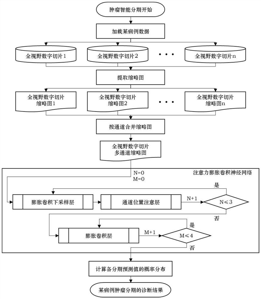
[0033] In this embodiment, intelligent staging diagnosis is performed on the full-field digital slices in the lung cancer database. The lung cancer database is a lung cancer database composed of 225 cases. There are 1071 full-field digital slices, of which 683 full-field digital slices are positive, and 388 The full-field digital slices were negative, that is, no tumor tissue was included. All full-field digital slices were cut into small pieces, a total of 161,973 pieces took up 92.2Gb of memory, and all full-field digital slices were extracted with thumbnails, a total of 1071 pieces took up 6.3Gb of memory.

[0034] The details of each step and the setting of model parameters are described in detail below.

[0035] Step 1, load case data: Traverse all files with the suffix .wsi format under the case path.

[0036] Use the os library in the python language to traverse the database of all cases to obtain the storage path of the cases. Use the sklearn library to divide 225 cas...


PUM


Abstract

The invention discloses a patient-level tumor intelligent diagnosis method based on full-view digital slicing; and the method comprises the following steps: obtaining a plurality of case databases ofa certain disease, and enabling each case database to name a folder after the ID of each patient, storing a plurality of full-view digital sections of biopsy tissue sections of all cases to be diagnosed and corresponding diagnosis results in each case database; extracting a digital slice with the minimum size at the bottom of the picture file stack of each full-view digital slice as a color imageof the full-view digital slice, and zooming the color image to obtain a color thumbnail; merging all the color thumbnails of a certain case into a full-view digital slice multichannel thumbnail according to channels; constructing a deep learning algorithm model; and loading all full-view digital slices of a certain case of the current disease, and outputting an intelligent diagnosis result. According to the invention, effective utilization of the full-view digital slice requiring large storage capacity is realized.

Description

technical field [0001] The invention relates to a patient-level tumor intelligent diagnosis method based on full-field digital slices. Background technique [0002] The full-view digital slice has billions of pixels, which takes up too much memory, and there will be insufficient memory when the computer processes it. The mainstream processing method is to cut the full-view digital slice into several small pieces, and input these small pieces into the algorithm in turn. The model is trained to obtain corresponding results. Three problems arise during this process. First, data preprocessing of full-field digital slices takes a lot of time. It takes about 3 minutes to cut a slice into small pieces, and cutting small pieces and training small pieces takes up a lot of computing resources. Secondly, a full-field digital slice can roughly cut out 280 512*512 tissue block pictures (after excluding blank blocks), and among them, there are very few key tissue blocks that provide im...

Claims


Application Information

Patent Timeline
Patent Type & Authority Applications(China)
IPC IPC(8): G16H50/20G16H30/40G06N3/04G06N3/08G06K9/62G06T3/40
CPCG16H50/20G16H30/40G06N3/08G06T3/4038G06T2200/32G06N3/045G06F18/253
Inventor 赵丹徐桂芝许铮铧
Owner HEBEI UNIV OF TECH
Learn More