Visual ultrasonic electrocoagulation hook

An ultrasonic electric and ultrasonic technology, which is used in medical science, heating surgical instruments, surgical instruments for suctioning substances, etc. problems such as surgical efficiency, to achieve the effect of shortening medical operation time, reducing the probability of secondary trauma, and improving medical operation efficiency

Pending Publication Date: 2021-01-26
FUJIAN MEDICAL UNIV UNION HOSPITAL
View PDF0 Cites 0 Cited by
  • Summary
  • Abstract
  • Description
  • Claims
  • Application Information

AI Technical Summary

Problems solved by technology

Electrocoagulation hooks and ultrasonic scalpels are one of the common equipment in laparoscopic surgery. However, with the continuous improvement of laparoscopic surgery requirements in the medical field, the deficiencies in the actual use of electrocoagulation hooks and ultrasonic scalpels need to be improved urgently.
[0003] At present, during clinical operations, medical staff often need to use the ultrasonic scalpel and electrocoagulation hook alternately to ensure the normal operation, but the frequent replacement of medical devices by doctors during the actual operation will not only interrupt the continuity of the operation, but also lead to the The prolongation of time will increase the potential safety hazards in the operation process and reduce the efficiency of medical operations. At the same time, the electric coagulation hook is not convenient for the doctor to perform multi-angle separation of the lesion tissue and the adjustment operation during resection, which reduces the doctor's surgical operation. Flexibility to reduce the effect of medical surgery

Method used


Image

  • Visual ultrasonic electrocoagulation hook
  • Visual ultrasonic electrocoagulation hook
  • Visual ultrasonic electrocoagulation hook

Examples


Embodiment Construction

[0026] The following examples are for illustrative purposes only and are not intended to limit the scope of the invention.

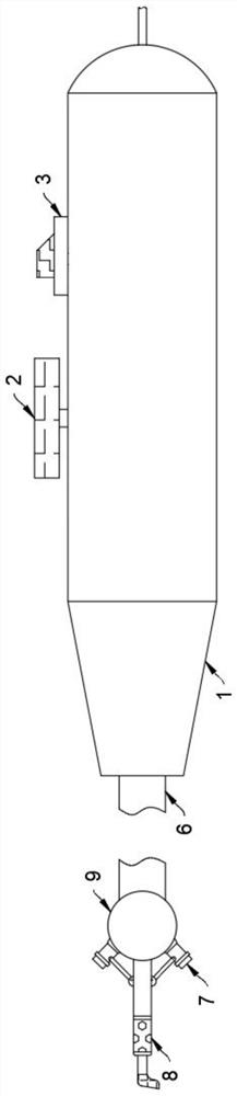
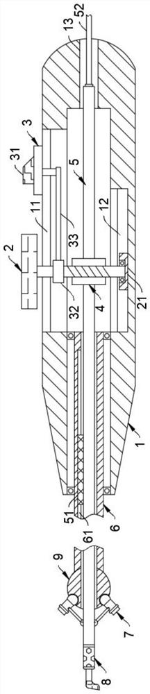
[0027] Such as Figure 1-5 As shown, a visual ultrasonic electrocoagulation hook includes a handle 1. It should be noted that the handle part of the handle 1 adopts a cylindrical structure, and the surface of the handle is provided with anti-skid lines. One end of the handle 1 adopts a tapered structure. The other end of 1 adopts a round head structure, which is convenient for doctors to hold and operate.

[0028] The handle 1 is rotatably connected to the adjustment wheel 2, and the adjustment wheel 2 is slidingly connected to the handle 1 through the push mechanism 3. It should be noted that the push mechanism 3 includes a push plate 31 and a rotating block 32. The push plate 31 adopts a trapezoidal structure, which is convenient for doctors to push Adjustment, the push plate 31 is fixedly connected with the rotating block 32 through the L-shaped guid...


PUM


Abstract

The invention belongs to the field of medical instruments and equipment, and particularly relates to a visual ultrasonic electrocoagulation hook. The visual ultrasonic electrocoagulation hook comprises a handle, wherein the handle is rotatably connected with an adjusting wheel; the adjusting wheel is slidably connected with the handle through a pushing mechanism; the adjusting wheel is rotatably connected with a catheter through a transmission mechanism; the end, close to the handle, of the catheter is slidably connected with a catheter sleeve; one end of the catheter sleeve is rotatably connected with the handle, and the other end of the catheter sleeve is slidably connected with an ultrasonic sensor; the ultrasonic sensor is movably connected with the catheter; and the end, away from thehandle, of the catheter is detachably connected with an electrocoagulation hook head. According to the visual ultrasonic electrocoagulation hook, by arranging the ultrasonic electrocoagulation hook with a visual effect, the situation that a doctor frequently replaces an ultrasonic knife and the electrocoagulation hook in the medical operation process is effectively avoided, the continuity of operation is improved, the medical operation efficiency is improved, the ultrasonic electrocoagulation hook is matched with the rotatable electrocoagulation hook head so that the doctor can conduct multi-angle separation and excision work on tissue at the focus, and the medical operation effect is improved.

Description

technical field [0001] The invention belongs to the field of medical equipment, and in particular relates to a visualized ultrasonic electrocoagulation hook. Background technique [0002] Laparoscopic surgery is a newly developed minimally invasive method, and it is an inevitable trend in the development of future surgical methods. With the rapid development of industrial manufacturing technology, the integration of related disciplines has laid a solid foundation for the development of new technologies and methods. Coupled with the increasingly skilled operation of doctors, many open operations in the past have now been replaced by endovascular surgery. Greatly increased surgical options. Electrocoagulation hooks and ultrasonic scalpels are one of the common equipment in laparoscopic surgery. However, as the requirements for laparoscopic surgery in the medical field continue to increase, the deficiencies in the actual use of electrocoagulation hooks and ultrasonic scalpels ...

Claims


Application Information

Patent Timeline
Patent Type & Authority Applications(China)
IPC IPC(8): A61B18/12A61B18/14A61B34/20A61B90/00
CPCA61B18/12A61B18/1482A61B2018/00607A61B2018/1422A61B2218/007A61B34/20A61B90/361A61B90/37A61B2034/2063A61B2034/2065A61B2090/378
Inventor 柴天赐杨晓杰张伟光陈椿康明强林江波
Owner FUJIAN MEDICAL UNIV UNION HOSPITAL
Why Eureka
Try Eureka