Artificial protein containing antigen-binding region of antibody and being fused with physiologically active peptide
A technology of physiological activity and binding activity, applied in the direction of fusion polypeptide, hybrid peptide, immunoglobulin, etc., can solve problems such as difficulties and loss of structural stability
- Summary
- Abstract
- Description
- Claims
- Application Information
AI Technical Summary
Problems solved by technology
Method used
Image
Examples
Embodiment 1
[0259] 1. Preparation of Peptide Fusion Proteins
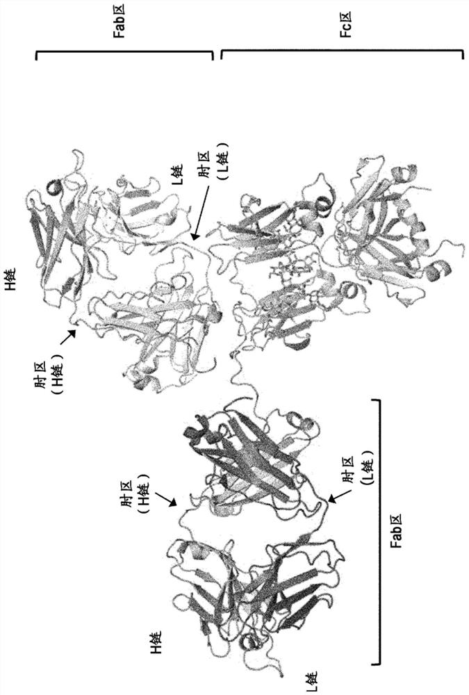
[0260] The DNA encoding the Fab region in the H chain of the human IgG1 monoclonal antibody against neuropilin 1 (N1 antibody, clone name YW64.3) and the DNA encoding the Hisx6 tag were incorporated into the expression vector pcDNA3.1 (Thermo Fisher Scientific company) (antibody H chain Fab region expression vector).
[0261] The amino acid sequence (SEQ ID NO: 2) of the N1 antibody H chain Fab region produced by this vector is shown in Figure 48 . It should be noted that the amino acid sequence (SEQ ID NO: 1) of the N1 antibody H chain is as Figure 47 show.
[0262] The DNA encoding the full-length region of the Lκ chain of the N1 antibody was incorporated into the expression vector pcDNA3.1 (antibody Lκ chain expression vector). but assembled in an unlabeled way. The amino acid sequence (SEQ ID NO: 3) of the N1 antibody Lκ chain produced by this vector is shown in Figure 49 .
[0263] As the physiologically activ...
Embodiment 2
[0298] 1. Preparation of Peptide Fusion Proteins
[0299] The peptide-fused IgG protein comprising the peptide-fused antibody H chain or the peptide-fused antibody L chain was obtained by combining an antibody H chain-peptide fusion vector with an unfused antibody Lκ chain expression vector, Or antibody L chain-peptide fusion vector and unfused antibody H chain IgG region expression vector co-expressed and secreted into its culture supernatant.
[0300] 40 µL of Protein A Sepharose (manufactured by GE Healthcare) was added to 0.25 mL of the recovered culture supernatant, and vortexed for 1 hour. Precipitate the Sepharose by centrifugation, remove the supernatant, wash the Sepharose twice with 1 mL of Tris-buffered saline (TBS, 20 mM Tris-HCl, 150 mM NaCl, pH 7.5), add 12.5 μL of TBS and 12.5 μL of SDS Add sample buffer for elution, and then heat at 95°C for 2 minutes to obtain a sample. 5 μL of the eluted sample was subjected to electrophoresis under reducing conditions an...
Embodiment 3
[0306] As the physiologically active peptide, aMD4 having the activity of binding to human Met receptor (Met) was used instead of mP6-9 having the activity of binding to human plexin B1, and the same procedure as in Example 1 was carried out.
[0307] aMD4 is a cyclic peptide having the amino acid sequence (SEQ ID NO: 34) of D-Tyr Arg Gln Phe Asn Arg Arg Thr His Glu Val Trp Asn LeuAsp Cys, and the chloroacetylated D-Tyr and Cys form a cyclic structure. The amino acid sequence (SEQ ID NO: 35) of aMD4 inserted into each site of the Fab region is Tyr Arg Gln Phe Asn Arg Arg ThrHis Glu Val Trp Asn Leu Asp.
[0308] It should be noted that, if Figure 24 and Figure 28 As shown in , Gly was used as a linker at the N-terminus and C-terminus of the inserted amino acid sequence (SEQ ID NO: 36-51).
[0309] The results are shown in Figure 25 ~ Figure 27 and Figure 29 ~ Figure 32 .
PUM
 Abstract
Description
Claims
Application Information
 - R&D
- Intellectual Property
- Life Sciences
- Materials
- Tech Scout
- Unparalleled Data Quality
- Higher Quality Content
- 60% Fewer Hallucinations
Browse by: Latest US Patents, China's latest patents, Technical Efficacy Thesaurus, Application Domain, Technology Topic, Popular Technical Reports.
© 2025 PatSnap. All rights reserved.Legal|Privacy policy|Modern Slavery Act Transparency Statement|Sitemap|About US| Contact US: help@patsnap.com


