
Ultrasonic thyroid nodule segmentation method based on asymmetric network

A thyroid nodule, asymmetric technology, applied in neural learning methods, biological neural network models, image analysis, etc., can solve the problems of extremely unbalanced foreground and background ratio, unable to obtain segmentation results, low image contrast, etc. Serious loss of image edge information, good accuracy and generalization ability, and the effect of solving too many interference factors

Pending Publication Date: 2021-02-19
JIANGNAN UNIV +1
View PDF0 Cites 3 Cited by
  • Summary
  • Abstract
  • Description
  • Claims
  • Application Information

AI Technical Summary

Problems solved by technology

However, for thyroid ultrasound images, the image contrast is low, the thyroid nodules are small, the ratio of foreground to background is extremely unbalanced, and there are many burrs on the border of nodules, which are easily interfered by tissues and organs around the thyroid gland, so good segmentation results cannot be obtained

Method used


Image

  • Ultrasonic thyroid nodule segmentation method based on asymmetric network
  • Ultrasonic thyroid nodule segmentation method based on asymmetric network
  • Ultrasonic thyroid nodule segmentation method based on asymmetric network

Examples


Embodiment Construction

[0022] The present invention will be further described in detail below in conjunction with the accompanying drawings and specific embodiments.

[0023] The present invention has constructed a kind of ultrasonic thyroid nodule method based on asymmetric network, and concrete steps are as follows:

[0024] (1) Cut the thyroid ultrasound image collected from the hospital into a single-channel grayscale image with a size of 256×256 to reduce the parameter amount of the network model. The data is divided into training set and test set according to 8:2. Due to the small number of data sets, a series of data enhancement operations such as flipping, rotating, and enhancing contrast are performed on the data.

[0025] (2) Construct an asymmetric network, send the processed data into the network for training, use Adam's gradient descent method for network optimization, and automatically adjust the learning rate to obtain the network model.

[0026] The steps of the second step of cons...


PUM


Abstract

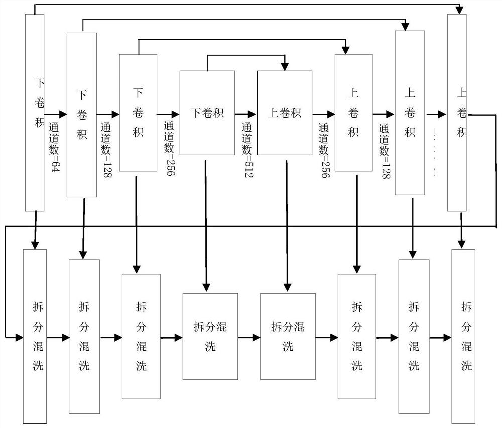
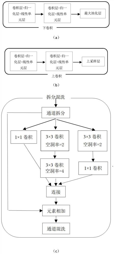
The invention discloses an ultrasonic thyroid nodule segmentation method based on an asymmetric network, and belongs to the field of deep learning image segmentation. The coding layer is a U-Net network, more context semantic feature information can be obtained, thyroid nodules with variable scales, shapes, positions and the like can be accurately detected, and the decoding layer uses the combination of hole convolution, residual connection and convolution kernel decomposition, so that detail textures and edge information can be gradually restored from high-dimensional global context semanticfeature information. According to the model constructed by the method, the segmentation precision similar to that of a high-precision model is achieved on a thyroid data set, the IOU is about 2% higher than that of a U-Net model, the time for averagely segmenting one picture is about 1.9 s, and the efficiency is greatly improved compared with the manual segmentation efficiency of doctors. The method solves the problems of excessive interference factors and serious loss of image edge information in a traditional method, and has good accuracy and generalization ability.

Description

technical field [0001] The invention belongs to the field of deep learning image segmentation, and in particular relates to a thyroid nodule ultrasound image segmentation method based on an asymmetric network. Background technique [0002] Thyroid disease is currently the most common nodular disease, and ultrasonography is the primary method for diagnosing thyroid nodules. However, the ultrasonic medical images collected by medical instruments have low contrast, more speckle noise, and thyroid nodules change in shape and position, which brings great difficulty to the diagnosis of thyroid diseases. [0003] In recent years, with the continuous development of medical imaging technology, computer-aided diagnosis has gradually attracted the attention of researchers. The use of neural networks to establish medical image segmentation models has been widely used, and the research on thyroid ultrasound image-aided diagnosis has also made great progress. In 2015, Ronneberger et al. ...

Claims


Application Information

Patent Timeline
IPC IPC(8): G06T7/12G06N3/04G06N3/08
CPCG06T7/12G06N3/084G06T2207/10132G06T2207/30096G06N3/048G06N3/045
Inventor 肖志勇吉淑滢柴志雷周锋盛丁炎张雨
Owner JIANGNAN UNIV
Learn More