A deep detection network to quantify the vascular morphological distribution of esophageal mucosal IPCLs

An esophageal mucosa, depth detection technology, applied in the field of medical image processing, can solve the problems of lack of quantifiable concepts, medical decision errors, visual fatigue, etc., to improve the efficiency of diagnosis, improve efficiency and accuracy, and reduce the amount of calculation.

Active Publication Date: 2022-07-22
FUDAN UNIV
View PDF0 Cites 0 Cited by
  • Summary
  • Abstract
  • Description
  • Claims
  • Application Information

AI Technical Summary

Problems solved by technology

In this case, clinicians need to observe all the structures, which is extremely easy to cause visual fatigue. Coupled with the lack of clinical experience, after observing 5-10 visual fields, clinicians often only remember the impression "Refer to Murphy's Law" for particularly profound parts, lacking an objective and quantifiable concept, which may easily lead to misjudgment of the condition and errors in medical decision-making

Method used


Image

  • A deep detection network to quantify the vascular morphological distribution of esophageal mucosal IPCLs
  • A deep detection network to quantify the vascular morphological distribution of esophageal mucosal IPCLs
  • A deep detection network to quantify the vascular morphological distribution of esophageal mucosal IPCLs

Examples


Embodiment Construction

[0029] The embodiments of the present invention will be described in detail below, but the protection scope of the present invention is not limited to the examples.

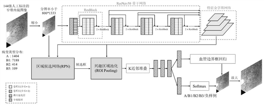
[0030] The present invention adopts figure 1 The network framework shown is trained using 144 narrow-band imaging endoscopic images that are collaboratively annotated by multiple experienced doctors, thereby obtaining a model that can automatically detect and diagnose esophageal squamous cell carcinoma foci from narrow-band imaging endoscopic images. The specific process is:

[0031] (1) Before training, the network parameters of the ResNet-50 model are randomly initialized, and the images in the training set are scaled so that the resolution does not exceed 800×1333, and the corresponding bounding box is also scaled at the same time. .

[0032] (2) During training, the image is first normalized according to mean=[0.485, 0.456, 0.406] and standard deviation=[0.229, 0.224, 0.225] to the three channels (R, G, B) ...


PUM


Abstract

The invention belongs to the technical field of medical image processing, in particular to a depth detection network for quantifying the morphological distribution of esophageal mucosal IPCLs blood vessels. The present invention includes feature extraction network and feature pyramid, region candidate network, interest region pooling and cluster distribution prior self-embedded cancer focus classification network, and visualization system on narrow-band imaging endoscopic images. The feature extraction network extracts the feature map of the input image; the feature pyramid fuses the features of different scales; the region candidate network proposes possible lesion regions; the pooling of the interest region pools the features to the suspicious lesion region; the cluster distribution prior self-embedding The cancer foci classification network of the system classifies the cancer foci; finally, it is visualized on the narrow-band imaging endoscopic image, and the cancer foci are box-marked with different colors. The present invention detects and diagnoses the cancer foci of early esophageal squamous cell carcinoma existing in the image, which can effectively improve the diagnosis efficiency and assist the doctor to obtain higher diagnosis accuracy.

Description

technical field [0001] The invention belongs to the technical field of medical image processing, and in particular relates to a depth detection network for quantifying the morphological distribution of esophageal mucosal IPCLs blood vessels. Background technique [0002] Esophageal cancer and gastric cancer have poor prognosis, with 5-year relative survival rates of 20.9% and 27.4%, respectively, bringing a serious burden to health care [11,13-14] . Standardized upper gastrointestinal cancer screening, treatment, and follow-up are effective means to reduce cancer morbidity and mortality. Among them, narrow-band imaging endoscopic screening is the first choice for the detection of upper gastrointestinal cancer. The pathological type and infiltration depth of esophageal mucosal lesions under narrow-band imaging endoscopy are mainly based on their unique vascular morphology of intrapapillary capillary loops (IPCLs). [0003] According to the classification criteria proposed ...

Claims


Application Information

Patent Timeline
Patent Type & Authority Patents(China)
IPC IPC(8): G06T7/00G06T3/40G06N3/08G06N3/04
CPCG06T7/0012G06T3/40G06N3/08G06T2207/20104G06T2207/30096G06T2207/20081G06T2207/10068G06N3/045
Inventor 钟芸诗颜波蔡世伦谭伟敏王沛晟李吉春阿依木克地斯·亚力孔
Owner FUDAN UNIV
Why Eureka
Try Eureka