Esophageal cancer lesion area search and identification modeling method based on evolutionary neural network structure

A neural network model and lesion area technology, applied in the field of esophageal cancer lesion area identification and modeling based on evolutionary neural network structure search, to achieve the effect of eliminating dependence, easy to use, and convenient to deploy

Active Publication Date: 2021-03-09
SICHUAN UNIV
View PDF8 Cites 7 Cited by
  • Summary
  • Abstract
  • Description
  • Claims
  • Application Information

AI Technical Summary

Problems solved by technology

[0006] In general: the convolutional neural network method has been widely used in the identification of esophageal cancer lesion

Method used


Image

  • Esophageal cancer lesion area search and identification modeling method based on evolutionary neural network structure
  • Esophageal cancer lesion area search and identification modeling method based on evolutionary neural network structure
  • Esophageal cancer lesion area search and identification modeling method based on evolutionary neural network structure

Examples


Embodiment Construction

[0049] In order to make the object, technical solution and advantages of the present invention clearer, the present invention is further described in detail. It should be understood that the specific embodiments described here are only used to explain the present invention, and are not intended to limit the present invention, that is, the described embodiments are only a part of the embodiments of the present invention, rather than all the embodiments.

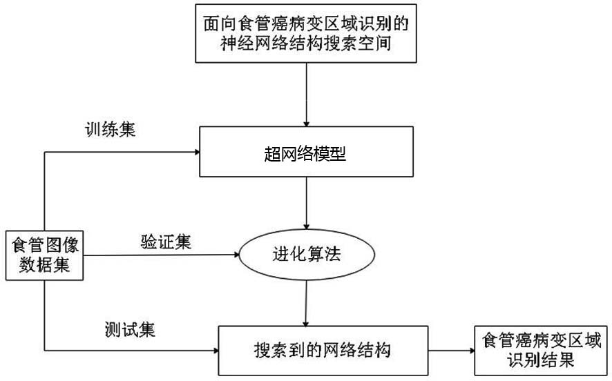
[0050] A method for identifying and modeling esophageal cancer lesion regions based on evolutionary neural network structure search, comprising the following steps:

[0051] S1: Collect and label the esophagus image data set used to train the neural network model:

[0052] Wherein, the esophageal image data set collected and marked for training the neural network model in step S1 includes the following steps:

[0053] S1-1: Record and collect video streams of esophageal endoscopy, screen and cut out video clips of NBI imaging m...


PUM


Abstract

The invention discloses an esophageal cancer lesion area search and identification modeling method based on evolutionary neural network structure, and relates to the technical field of image mode recognition and medical images, and the method comprises the following steps: S1, collecting and labeling an esophageal image data set for training a neural network model; S2, constructing a neural network structure search space for esophageal cancer lesion area identification; S3, training a super-network model for esophageal cancer lesion area identification; S4, searching an optimal neural networkstructure on the constructed super-network model by using an evolutionary algorithm; and S5, finely adjusting the searched neural network structure, and predicting a lesion area on the newly input esophageal image. The dependence of neural network structure design on expert experience in an esophageal cancer intelligent recognition task is eliminated, so that the deep neural network method is easier to use in the aspect of esophageal cancer lesion area recognition.

Description

technical field [0001] The invention relates to the technical field of image pattern recognition and medical images, in particular to a method for identifying and modeling esophageal cancer lesion regions based on evolutionary neural network structure search. Background technique [0002] Esophageal cancer (Esophageal Cancer, EC) is a malignant disease with a high mortality rate [A. K. Rustgiand H. B. El-Serag, “Esophageal cancer,” New England Journal of Medicine, vol. 371, no. 26, pp. 2499–2509 , 2014.], studies have shown that its 5-year survival rate is less than 20% [ X.-X.Chen, Q. Zhong, Y. Liu, S.-M. Yan, Z.-H. Chen, S.-Z . Jin, T.-L. Xia, R.-Y.Li, A.-J. Zhou, Z. Su et al., “Genomic comparison of esophageal squamous cellcarcinoma and its precursor lesions by multi-region whole-exome sequencing ,” Nature Communications, vol. 8, no. 1, pp. 1–12, 2017.]. Gastroscopy has been widely used in the diagnosis of early esophageal cancer, which can provide guidance for early in...

Claims


Application Information

Patent Timeline
IPC IPC(8): G06F30/27G06T7/00G06T3/40G06N3/04G06N3/08G06F111/04G06F111/06G06F111/08
CPCG06F30/27G06T7/0012G06T3/4007G06N3/08G06F2111/04G06F2111/06G06F2111/08G06T2207/20081G06T2207/20084G06T2207/30096G06N3/045
Inventor 章毅胡兵张潇之周尧刘伟吴雨袁湘蕾
Owner SICHUAN UNIV
Why Eureka
Try Eureka