Fundus optic disc and fovea centralis real-time detection device and method based on deep learning

A technology of deep learning and detection method, which is applied in the field of medical image processing to achieve the effects of improving detection accuracy, simplifying processing process and shortening detection time.

Pending Publication Date: 2021-03-23
TIANJIN UNIV
View PDF4 Cites 1 Cited by
  • Summary
  • Abstract
  • Description
  • Claims
  • Application Information

AI Technical Summary

Problems solved by technology

However, these methods still have some room for improvement in terms of multi-scale feature extraction and operation speed.

Method used


Image

  • Fundus optic disc and fovea centralis real-time detection device and method based on deep learning
  • Fundus optic disc and fovea centralis real-time detection device and method based on deep learning
  • Fundus optic disc and fovea centralis real-time detection device and method based on deep learning

Examples


Embodiment Construction

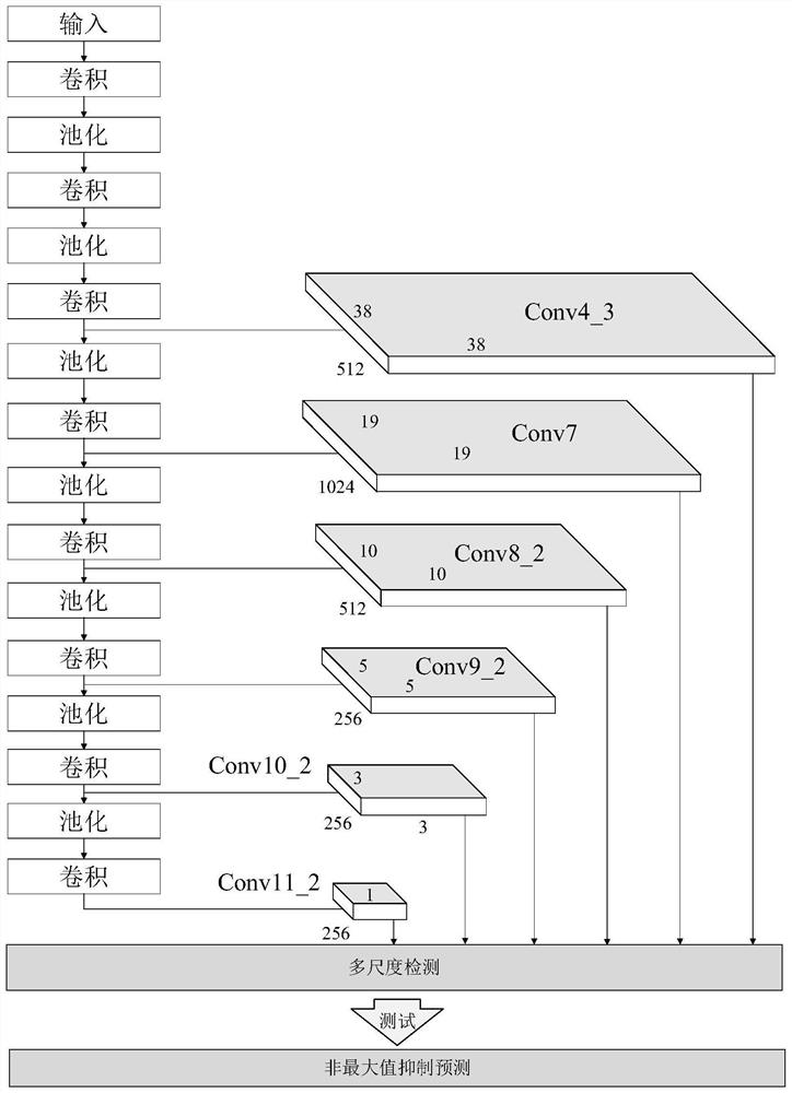
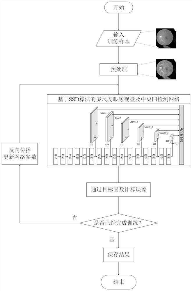
[0036] The invention aims to realize the real-time detection of the optic disc and fovea of ​​the fundus based on deep learning, and improve the detection accuracy and speed of the optic disc and fovea. The present invention builds a fundus optic disc and fovea detection network based on the single-stage multi-frame detection (Single Shot MultiBoxDetector, SSD) algorithm, and optimizes the parameters of the network model through multiple iterations to realize the detection of the optic disc and fovea center in a large number of fundus images. Real-time positioning. Method steps of the present invention are as follows:

[0037] Step 1: Construct training data set and test data set respectively;

[0038] Step 2: Build a multi-scale fundus optic disc and fovea detection network model based on the SSD algorithm;

[0039] Step 3: Input the training set into the network, get predictions through forward propagation and compare them with expert annotations, and optimize network para...


PUM


Abstract

The invention belongs to the technical field of medical image processing, and aims to realize real-time fundus optic disc and fovea centralis detection based on deep learning and improve the detectionprecision and speed of the optic disc and the fovea centralis. Therefore, according to the technical scheme, the fundus optic disc and fovea centralis real-time detection device and method based on deep learning are adopted, a fundus optic disc and fovea centralis detection network based on a single-stage multi-frame detection SSD algorithm is constructed, parameters of a network model are optimized through multiple times of iterative learning, and the detection precision of the network model is improved. And real-time positioning of the optic disc and the fovea centralis center in a large number of fundus images is realized. The method is mainly applied to medical image processing occasions.

Description

technical field [0001] The invention belongs to the technical field of medical image processing, and relates to a method for detecting an optic disc and a fovea of ​​a fundus image based on deep learning. Background technique [0002] With the rapid development of computer technology and people's increasing concern for health, computer-aided diagnosis technology has been more and more widely used in various medical fields. Computer-aided diagnosis technology mainly uses computers to process and analyze medical information, including medical data processing, medical image enhancement, medical image segmentation, important tissue area detection, etc., which has important reference significance for medical personnel's diagnosis and decision-making. Among them, medical image processing technology is an important part of computer-aided diagnosis, and is currently widely used in the fields of lesion region extraction and specific tissue measurement. [0003] In ophthalmology, the...

Claims


Application Information

Patent Timeline
IPC IPC(8): G06T7/00G06N3/04G06N3/08
CPCG06T7/0004G06N3/08G06T2207/10004G06N3/045
Inventor 唐晨谢慧颖
Owner TIANJIN UNIV
Why Eureka
Try Eureka