Intracranial hemorrhage CT image segmentation method based on deep learning

A technology of intracranial hemorrhage and CT images, which is applied in the field of artificial intelligence and medical image processing, can solve the problems of excessive differences in hemorrhage areas and the impact of segmentation, and achieve the effects of assisting diagnosis, avoiding errors, and meeting basic clinical needs

Active Publication Date: 2021-04-06
XIANGTAN UNIV
View PDF8 Cites 6 Cited by
  • Summary
  • Abstract
  • Description
  • Claims
  • Application Information

AI Technical Summary

Problems solved by technology

[0003] In order to improve the deficiencies of the existing CT image segmentation technology of intracranial hemorrhage and solve the impact of excessive bleeding area differences on segmentation, the present invention provides a CT image segmentation method of intracranial hemorrhage based on deep learning

Method used


Image

  • Intracranial hemorrhage CT image segmentation method based on deep learning
  • Intracranial hemorrhage CT image segmentation method based on deep learning

Examples


Embodiment Construction

[0022] The present invention will be further described in detail below in conjunction with the accompanying drawings and specific embodiments.

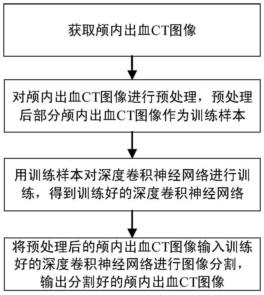
[0023] Such as figure 1 As shown, the present invention provides a method for segmenting CT images of intracranial hemorrhage based on deep learning, specifically comprising the following steps:

[0024] 1) Obtain CT images of intracranial hemorrhage;

[0025] 2) Preprocess the CT images of intracranial hemorrhage. Since the CT image data of intracranial hemorrhage used are collected from different imaging devices in different hospitals, the images of different cases have inconsistent sizes. It is necessary to adjust the CT images of intracranial hemorrhage to conform to the network model input. Preset size, the process is: for the acquired CT images of intracranial hemorrhage, adjust the size of each CT image of intracranial hemorrhage according to the preset image size, fill the edge with 0 pixels for images that are less than the ...


PUM


Abstract

The invention provides an intracranial hemorrhage CT image segmentation method based on deep learning. The intracranial hemorrhage CT image segmentation method comprises the steps of obtaining an intracranial hemorrhage CT image; preprocessing the intracranial hemorrhage CT image, and taking part of the preprocessed intracranial hemorrhage CT image as a training sample; training the deep convolutional neural network by using the training sample to obtain a trained deep convolutional neural network; and inputting the preprocessed intracranial hemorrhage CT image into the trained deep convolutional neural network for image segmentation, outputting the segmented intracranial hemorrhage CT image, and displaying a hemorrhage region segmentation result of the intracranial hemorrhage CT image through a GUI interface. The high-level features of the image are automatically extracted by means of the deep convolutional neural network, and the bleeding area is segmented, so that the problem of data imbalance caused by overlarge difference of the bleeding area is effectively solved, and high-precision segmentation is realized.

Description

technical field [0001] The present invention relates to the fields of artificial intelligence and medical image processing, in particular to a deep learning-based CT image segmentation method for intracranial hemorrhage. Background technique [0002] Intracranial hemorrhage (ICH) refers to the bleeding caused by the rupture of blood vessels in the brain. The hematoma can compress the surrounding nerve tissue and induce functional impairment. Intracranial hemorrhage may be caused by traffic accidents, trauma, high blood pressure, vascular lesions, brain tumors, etc. This disease has become a common disease. It is extremely important for patients to diagnose and give treatment options. With the development of imaging technology, the clinical diagnosis of intracranial hemorrhage is mainly for radiologists to examine computerized tomography (CT) images to detect and locate intracranial hemorrhage areas. Due to the low contrast of the image and the blurred boundary of the hemor...

Claims


Application Information

Patent Timeline
Patent Type & Authority Applications(China)
IPC IPC(8): G06T7/11G06N3/04G06N3/08
CPCG06T7/11G06N3/08G06T2207/10081G06T2207/20081G06T2207/30016G06T2207/30101G06N3/045
Inventor 胡凯侯媛媛张园高协平
Owner XIANGTAN UNIV
Why Eureka
Try Eureka