Ultra-thin small-caliber high-resolution endoscopic microscope objective

A microscope objective, high-resolution technology, used in microscopes, telescopes, optics, etc., can solve problems such as narrow working clamps, delayed treatment, patient pain, etc., and achieve high-resolution processing, improved resolution, and good processability. Effect

Active Publication Date: 2021-04-09
ZHEJIANG LAB +1
View PDF9 Cites 0 Cited by
  • Summary
  • Abstract
  • Description
  • Claims
  • Application Information

AI Technical Summary

Problems solved by technology

However, for narrow spaces, such as narrow areas of the biliopancreatic duct, high-resolution endoscopes cannot be used, and the doctor may not get cancerous tissue after taking more than 5 samples, resulting in missed detection and delayed treatment.
In addition, the method of in vitro biopsy takes about two weeks. At this stage, the patient's condition may develop rapidly, which is very unfavorable to the patient's treatment. Moreover, endoscopic treatment after the pathological diagnosis of the lesion by routine biopsy will delay Post-treatment time, increasing the patient's pain and psychological and medical burden
This will not only bring pain to the patient and increase the risk of lesion spread, but also require a long wait for pathological test results, resulting in low diagnosis and treatment efficiency
Moreover, the working channel of the traditional endoscope is very narrow, and its diameter is between 1.2mm-4.2mm

Method used


Image

  • Ultra-thin small-caliber high-resolution endoscopic microscope objective
  • Ultra-thin small-caliber high-resolution endoscopic microscope objective
  • Ultra-thin small-caliber high-resolution endoscopic microscope objective

Examples


Embodiment 1

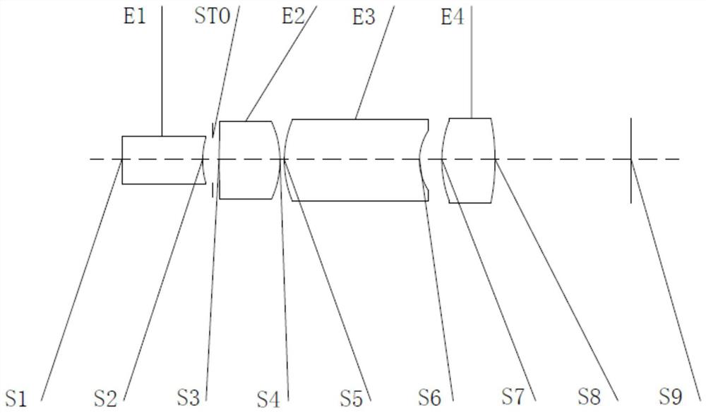
[0101] The surface type, curvature radius, thickness, semi-diameter and material of each lens in this embodiment are shown in Table 1.

[0102] Table 1 Embodiment 1 Endoscopic microscope objective lens parameters

[0103]

[0104]

Embodiment 2

[0106] The surface type, curvature radius, thickness, semi-diameter and material of each lens in this embodiment are shown in Table 2.

[0107] Table 2 Embodiment 2 endoscopic microscope objective lens parameters

[0108] face number surface type Radius of curvature (mm) Thickness (mm) Semi-aperture(mm) Material OBJ sphere endless 0.1511 0.1760 S1 sphere endless 0.5748 0.1707 1.50,66.2 S2 sphere 0.5734 0.0721 0.1565 STO sphere endless 0.0454 0.1620 S3 sphere 3.7036 0.4341 0.1842 1.69,21.2 S4 sphere -0.6595 0.0300 0.2711 S5 sphere 0.7519 0.9625 0.2909 1.75,35.0 S6 sphere 0.3889 0.1592 0.2126 S7 sphere 0.8001 0.3795 0.2782 1.75,35.0 S8 sphere -1.5341 0.9690 0.3000 S9 sphere endless 0.3010

Embodiment 3

[0110] The surface type, curvature radius, thickness, semi-diameter and material of each lens in this embodiment are shown in Table 3.

[0111] Table 3 Embodiment 3 endoscopic microscope objective lens parameters

[0112]

[0113]


PUM


Abstract

The invention relates to an ultra-thin small-caliber high-resolution endoscopic microscope objective which comprises a first lens, a diaphragm, a second lens, a third lens and a fourth lens sequentially arranged from the object side. All the lenses are spherical lenses and are separately arranged by taking air as an interval, the first lens has negative focal power, the object side surface of the first lens is a plane, and the image side surface of the first lens is a concave surface; the second lens has positive focal power, the object side surface of the second lens is a convex surface, and the image side surface is a convex surface;the third lens has positive focal power, the object side surface of the third lens is a convex surface, and the image side surface of the third lens is a concave surface; the fourth lens has positive focal power, the object side surface of the fourth lens is a convex surface, the image side surface of the fourth lens is a convex surface, the maximum clear aperture is less than or equal to 0.6, the axial distance from the object side surface of the first lens to the imaging surface is less than or equal to 4.0 mm, and 1.1< f*TAN(HFOV)/T34< 2.3. The endoscopic microscope objective is small in caliber, small in thickness, high in resolution, compact in structure, convenient to machine and install and good in imaging quality, and the requirements for miniaturization and light weight of the medical endoscopic microscope objective are met.

Description

technical field [0001] The invention relates to the technical field of endoscopic microscopic objective lenses, in particular to an ultra-thin small-caliber high-resolution endoscopic microscopic objective lens. Background technique [0002] With the continuous development of medical technology, medical endoscopes are often used in clinical diagnosis and treatment. The probe-type confocal microendoscope can send the probe to the designated part through the natural channel of the human body, and obtain local histological images to realize the accurate diagnosis of small lesions, gastrointestinal lesions and early gastrointestinal cancer. Traditional endoscopic microscope objectives can only provide tissue surface image information, and due to the limitation of human body structure, the narrow area cannot be reached by routine endoscopy, and biopsy is often required for further diagnosis. However, the current in vitro biopsy method is random inspection. The general operation ...

Claims


Application Information

Patent Timeline
Patent Type & Authority Applications(China)
IPC IPC(8): G02B23/24G02B13/00G02B21/02
CPCG02B23/243G02B13/005G02B21/02
Inventor 游兴海杨青王立强徐璟罡董震宇
Owner ZHEJIANG LAB
Why Eureka
Try Eureka