Multi-scale DC-CUNets liver tumor segmentation method based on bottleneck structure

A liver tumor, multi-scale technology, applied in the field of medical image processing, can solve the problem of insufficient segmentation accuracy of liver tumors, and achieve the effect of improving the overall segmentation accuracy, optimizing the training process, and reducing the probability of false negatives and false positives.

Active Publication Date: 2021-04-27
NANJING UNIV OF POSTS & TELECOMM
View PDF2 Cites 1 Cited by
  • Summary
  • Abstract
  • Description
  • Claims
  • Application Information

AI Technical Summary

Problems solved by technology

[0003] In order to solve the problem of insufficient segmentation accuracy of liver tumors in the prior art, the present invention proposes a multi-scale DC-CUNets liver tumor segmentation method based on the bottleneck s

Method used


Image

  • Multi-scale DC-CUNets liver tumor segmentation method based on bottleneck structure
  • Multi-scale DC-CUNets liver tumor segmentation method based on bottleneck structure
  • Multi-scale DC-CUNets liver tumor segmentation method based on bottleneck structure

Examples


Embodiment Construction

[0065] Below in conjunction with accompanying drawing, technical scheme of the present invention will be further described:

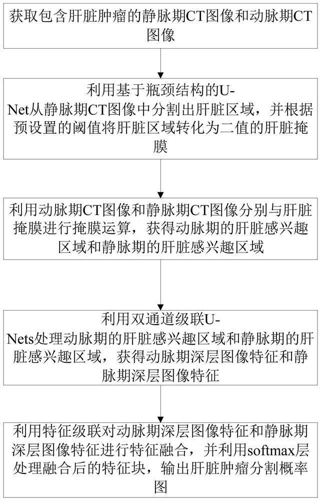
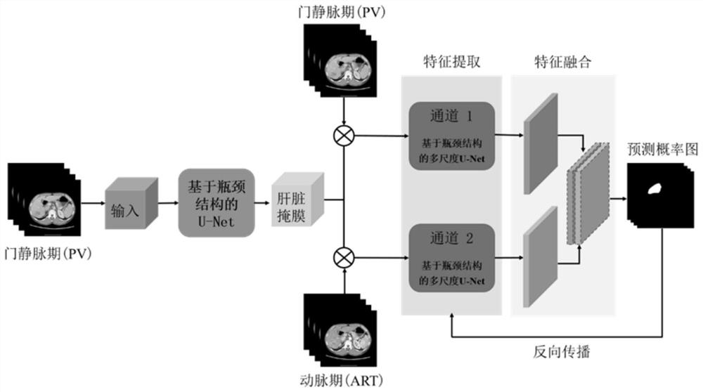
[0066] The present invention proposes a multi-scale DC-CUNets liver tumor segmentation method based on the bottleneck structure, which uses dual-channel U-Nets to jointly process the venous phase and arterial phase images of enhanced CT, and solves the problem of multi-scale DC-CUNets in the prior art. The problem of fusion of early image features, the problem of the scale of liver tumors, and the optimization of the network training process, such as figure 1 , 2 shown, including the following steps:

[0067] Step 1. Obtain a CT image in the venous phase and a CT image in the arterial phase including the liver tumor;

[0068] Step 2. Use the U-Net based on the bottleneck structure to segment the liver region from the venous phase CT image, and convert the liver region into a binary liver mask according to the preset threshold;

[0069] Step 3, using ...


PUM


Abstract

The invention discloses a multi-scale DC-CUNets liver tumor segmentation method based on a bottleneck structure, and aims to solve the technical problem of insufficient liver tumor segmentation precision in the prior art. The method comprises the following steps: obtaining a liver mask from a vein CT image by utilizing U-Net based on a bottleneck structure; performing mask operation on the CT images in the arterial phase and the venous phase and the liver mask to obtain liver regions of interest in the arterial phase and the venous phase; processing liver regions of interest in the arterial phase and the venous phase by using dual-channel cascade U-Nets to obtain deep image features in the arterial phase and the venous phase; and performing feature fusion on the deep image features in the arterial phase and the venous phase, processing the fused feature blocks by using a softmax layer, and outputting a liver tumor segmentation probability graph. According to the method, liver tumor segmentation can be rapidly and accurately carried out.

Description

technical field [0001] The invention relates to a multi-scale DC-CUNets liver tumor segmentation method based on a bottleneck structure, and belongs to the technical field of medical image processing. Background technique [0002] As a common malignant tumor disease, liver cancer is one of the most common cancers in the world. Accurate diagnosis of liver tumors based on CT effects has very important guiding significance for liver cancer research, clinical diagnosis and surgical treatment. At present, more and more studies have used computer image processing technology as an auxiliary diagnosis and treatment method to segment liver and tumors from CT images. However, information such as liver size and location varies from person to person, and intraperitoneal Due to the complex connection of organs and tissues in medical imaging, the low contrast of medical images, the diversity and spread of tumors, the blurred boundaries of liver and tumors, and the uneven density distribut...

Claims


Application Information

Patent Timeline
IPC IPC(8): G06T7/11G06T7/136G06K9/32G06K9/46G06K9/62G06N3/04G06N3/08
CPCG06T7/11G06T7/136G06N3/08G06T2207/10081G06T2207/30056G06T2207/30096G06V10/25G06V10/40G06N3/045G06F18/253
Inventor 胡栋徐畅畅庞雨薇
Owner NANJING UNIV OF POSTS & TELECOMM
Why Eureka
Try Eureka