Anti-CD123 antibody and application thereof

An antibody and antigen technology, applied in the field of immunology, can solve the problems of poor specificity and low affinity of CD123 antibody, and achieve the effect of strong binding specificity

Active Publication Date: 2021-05-28
INST OF HEMATOLOGY & BLOOD DISEASES HOSPITAL CHINESE ACADEMY OF MEDICAL SCI & PEKING UNION MEDICAL COLLEGE +1
View PDF3 Cites 4 Cited by
  • Summary
  • Abstract
  • Description
  • Claims
  • Application Information

AI Technical Summary

Problems solved by technology

[0007] One aspect of the present invention is aimed at the problems of low affinity and poor specific

Method used


Image

  • Anti-CD123 antibody and application thereof
  • Anti-CD123 antibody and application thereof
  • Anti-CD123 antibody and application thereof

Examples


Embodiment 1

[0123] Example 1: Screening of Mouse Hybridoma Monoclonal Antibodies

[0124] Using human CD123 protein as the immunogen, Balb / c mice were immunized by intraperitoneal injection, booster immunization was carried out at the 3rd week and the 5th week after the initial immunization, and the mouse tail blood was collected on the 8th day after the booster immunization , stand at room temperature for 1 hour, centrifuge at 12,000 rpm for 10 minutes at 4°C, collect serum, and dilute with PBS to different concentrations: 1:200, 1:400, 1:800, 1:1600, 1:3200, 1:6400, 1 : 12800. Collect THP-1 cells, wash once with PBS, count the cells, use 1×10 for each sample 6 Add 100 μl of serum with different dilutions to the cells, and in the negative control group, replace the antiserum with the serum of unimmunized mice, incubate at 4°C for 1 hour, wash twice with PBS; add 2 μl of PE-labeled rat anti-mouse IgG antibody , and incubate at 4°C in the dark for 40 minutes; cells were resuspended in 50...

Embodiment 2

[0126] Embodiment 2: Ascites preparation and purification

[0127] Wash hybridoma cells with sterile PBS solution, 5x10 6 The cell volume of 0.5ml / mouse was intraperitoneally injected into the liquid paraffin presensitized Balb / c mice. Collect ascites after 7 to 10 days, room temperature 3000rpm, 10min, collect supernatant. The antibody was crudely purified with saturated ammonium sulfate with a final concentration of 33%. The method was to take 1 part of ascitic fluid, add 1 part of PBS, add 1 part of saturated ammonium sulfate dropwise, stir while adding, overnight at 4°C, and centrifuge at 10,000 rpm for 10 minutes to remove the supernatant , Dissolve the precipitate with a small amount of PBS, dialyze with PBS for 24 hours at 4°C to desalt, and change the medium 3 times during this period. According to the purification manual provided by GE, the purified antibody was further purified by using the AKTA protein purification system and 1ml Protein G purification prepacked c...

Embodiment 3

[0128] Example 3: Monoclonal antibody titer detection

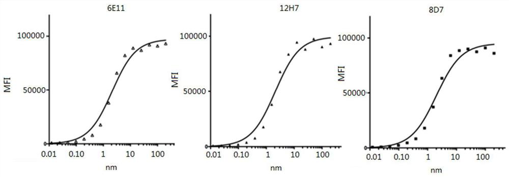
[0129] Fluorescently label the pure antibody, PE directly labeled with 6E11, 8D7, 12H7 antibody. Labeled antibodies at final concentrations of 400nM, 200nM, 100nM, 50nM, 25nM, 12.5nM, 6.25nM, 3.2nM, 1.6nM, 0.8nM, 0.4nM, 0.2nM, 0.1nM, 0.05nM, 0.025nM, 0.0125nM, 0.0061nM, 0.003nM and 2.5×10 5 3T3 (transfected with CD123) were incubated at room temperature for 30 min, and protected from light. Centrifuge at 1800 rpm for 10 min, discard the supernatant, wash the cells with PBS, repeat three times, resuspend the cells in 400 μl of PBS, measure the fluorescence intensity by FACS, and calculate the Mean value. The Kd value of the antibody was calculated with the data analysis software GraphPad Prism 5. The result is as figure 1 As shown, the Kd values ​​of 12H7, 6E11, and 8D7 are 2.09×10 -9 M, 2.10×10 -9 M, 2.01×10 -9 M.


PUM


Abstract

The invention discloses an antibody specifically binding to CD123. The antibody comprises: a) CDR1, CDR2 and CDR3 regions of a heavy-chain variable region shown as SEQ ID No. 1 to 3 or regions 80% or more homogeneous with the CDR1, CDR2 and CDR3 regions; or b) CDR1, CDR2 and CDR3 regions of a heavy-chain variable region shown as SEQ ID No. 4 to 6 or regions 80% or more homogeneous with the CDR1, CDR2 and CDR3 regions; or c) CDR1, CDR2 and CDR3 regions of a heavy-chain variable region shown as SEQ ID No. 7 to 9 or regions 80% or more homogeneous with the CDR1, CDR2 and CDR3 regions. The monoclonal antibody can be used for detecting cells expressing CD123, and can also be applied to tumor immunotherapy independently or be applied to tumor immunotherapy in combination with other methods; that is, the monoclonal antibody can be effectively applied to preparation of drugs for treating tumors, infectious diseases and autoimmune diseases, resisting immunological rejection and the like.

Description

technical field [0001] The present invention relates to the field of immunology, more specifically, the present invention relates to anti-CD123 antibody and application thereof. Background technique [0002] LSCs are the first cancer stem cells confirmed by Lapidot et al. It has similar unlimited proliferation and self-renewal capabilities to normal hematopoietic stem cells, most of them are in the quiescent phase, have self-protection mechanisms and survive in a highly protective microenvironment. Being able to escape the killing of conventional chemotherapy drugs, the persistence of LSCs is considered to be the root cause of leukemia occurrence, relapse, and drug resistance. Effective removal of LSCs has become an important goal of leukemia treatment. [0003] In addition to having the same surface antigen phenotype as normal hematopoietic stem cells, AML HSCs (CD34 + , CD38 - , CD71 - 、HLA - DR - ), there are some self-specific surface antigen phenotypes (CD90 - / CD...

Claims


Application Information

Patent Timeline
IPC IPC(8): C07K16/28C12N15/13A61K39/395A61P35/00A61P31/00A61P37/00A61P37/06
CPCC07K16/2866A61P35/00C07K2317/565A61K2039/505A61P35/02G01N33/574C07K2317/92
Inventor 熊冬生卢杨范冬梅王建祥张砚君叶舟张益枝杨纯正
Owner INST OF HEMATOLOGY & BLOOD DISEASES HOSPITAL CHINESE ACADEMY OF MEDICAL SCI & PEKING UNION MEDICAL COLLEGE
Why Eureka
Try Eureka