Carotid artery ultrasound image blood vessel and intima positioning method based on deep learning network

A deep learning network and ultrasound image technology, applied in the field of medical image analysis, can solve problems such as dependence on accuracy, difficulty in ensuring real-time algorithm performance, and lack of automation, achieving great value and meaning, and easy real-time calculation.

Pending Publication Date: 2021-06-15
成都思多科医疗科技有限公司
View PDF0 Cites 1 Cited by
  • Summary
  • Abstract
  • Description
  • Claims
  • Application Information

AI Technical Summary

Problems solved by technology

The above carotid artery LII and MAI segmentation methods mainly have two shortcomings: first, they are not fully automated, and still require manual intervention by doctors, and second, both dynamic programming and Snake methods rely on the accuracy of the initial contour acquisition.

Method used


Image

  • Carotid artery ultrasound image blood vessel and intima positioning method based on deep learning network
  • Carotid artery ultrasound image blood vessel and intima positioning method based on deep learning network
  • Carotid artery ultrasound image blood vessel and intima positioning method based on deep learning network

Examples


Embodiment 1

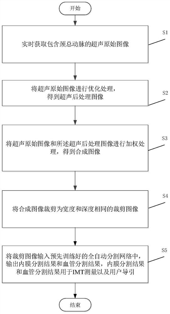
[0058] A flow chart of a carotid artery ultrasound image vessel and intima positioning method based on deep learning network image 3 As shown, it mainly includes the following steps:

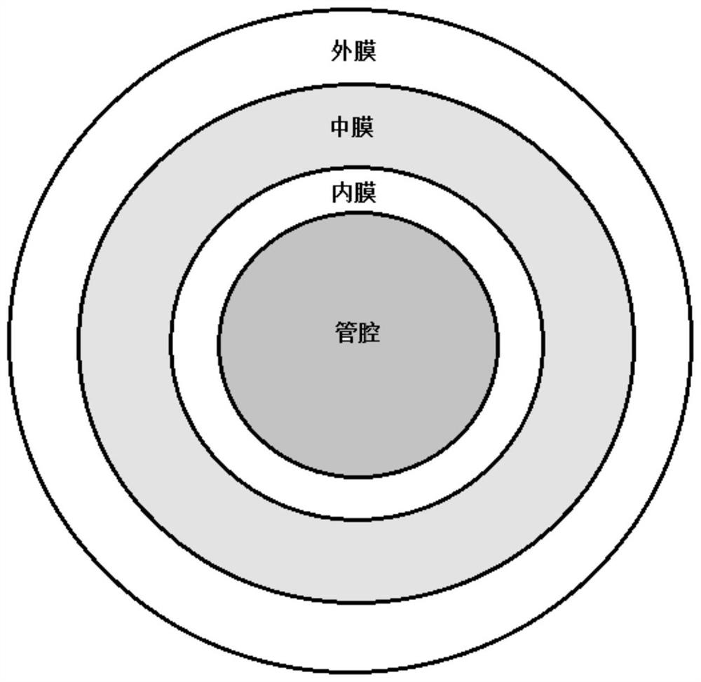
[0059] S1, real-time acquisition of original ultrasonic images including the common carotid artery;

[0060] S2. Optimizing the original ultrasonic image to obtain an ultrasonic post-processing image;

[0061] S3, performing weighting processing on the original ultrasonic image and the ultrasonic post-processing image to obtain a weighted image;

[0062] S4, cropping the weighted image into a cropped image with the same width and depth;

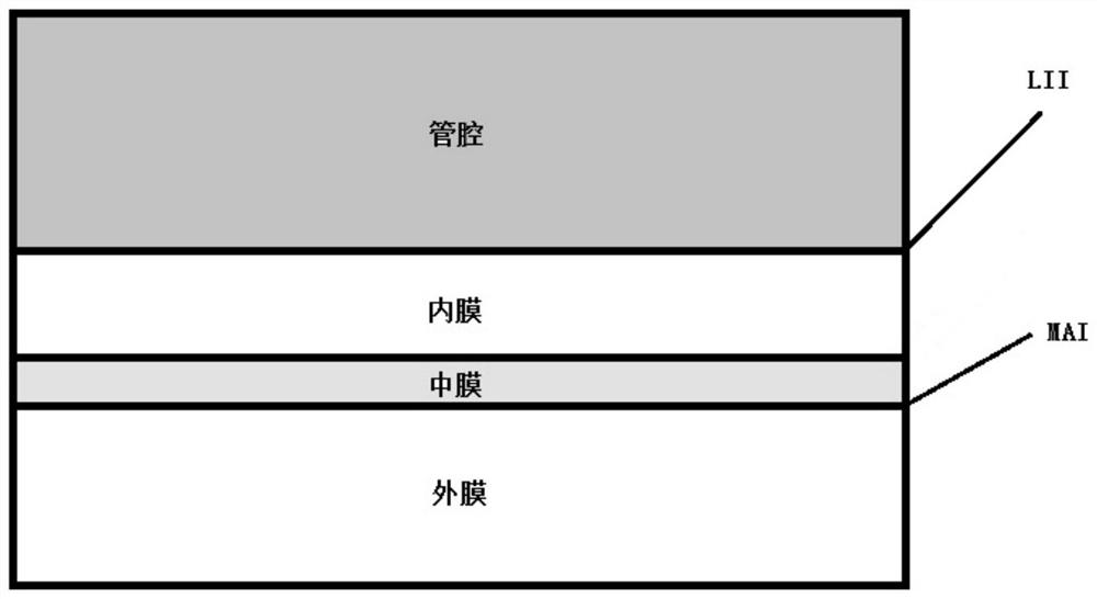
[0063] S5, input the cropped image into the pre-trained automatic segmentation network, output the intima segmentation result and blood vessel segmentation result, and use the intima segmentation result and blood vessel segmentation result for IMT measurement and user guidance.

[0064] Wherein, the schematic flowchart of obtaining the cropped image by step...


PUM


Abstract

The invention relates to the field of medical image analysis, in particular to a carotid artery ultrasound image blood vessel and intima positioning method based on a deep learning network. The method comprises the following steps: S1, acquiring an ultrasonic original image containing the common carotid artery in real time; S2, performing optimization processing on the ultrasonic original image to obtain an ultrasonic post-processing image; S3, weighting the ultrasonic original image and the ultrasonic post-processing image to obtain a weighted image; S4, cutting the weighted image into cut images with the same width and depth; and S5, inputting the cut image into a pre-trained full-automatic segmentation network, outputting an intima segmentation result and a blood vessel segmentation result, and applying the intima segmentation result and the blood vessel segmentation result to IMT measurement and user guidance. Accurate positioning and segmentation of blood vessels and intima of blood vessels are achieved. Compared with a traditional algorithm, the method does not need manual intervention at all, and compared with a secondary structure, real-time calculation is easier to achieve, and the real-time requirement is met.

Description

technical field [0001] The present invention relates to the technical field of medical image analysis, in particular to a carotid artery ultrasonic image blood vessel and intima positioning method based on a deep learning network. Background technique [0002] The basic pathology of cardiovascular and cerebrovascular diseases (CVD) is atherosclerosis, and carotid intimal thickness (cIMT) is an important indicator to measure the degree of atherosclerosis. Clinically, after the ultrasound images of the carotid artery are usually collected using non-destructive ultrasound imaging technology, the sonographer usually manually marks the lumen-intima boundary (LII) and adventitia-media on the collected ultrasound images of the common carotid artery Boundary (MAI), to achieve the measurement of IMT. [0003] In the ultrasound image of the carotid artery, the proximal vessel wall is represented by the outer ring, the lumen is located in the middle of the image, and the distal vessel...

Claims


Application Information

Patent Timeline
IPC IPC(8): G06T7/00G06T7/11G06T7/13G06T7/187G06T5/00G06K9/32G06K9/62G06N3/04G06N3/08
CPCG06T7/0012G06T7/11G06T7/13G06T7/187G06T5/007G06N3/08G06T2207/10132G06T2207/20081G06T2207/20084G06T2207/20104G06T2207/30101G06V10/25G06N3/047G06N3/045G06F18/214
Inventor 张琳刘西耀高君
Owner 成都思多科医疗科技有限公司
Why Eureka
Try Eureka