
Automatic precise positioning scanning method of PET/CT system

A scanning method and precise positioning technology, applied in computerized tomography scanners, patient positioning for diagnosis, medical science, etc., can solve the problems of tissue cell damage, radiation hazards, etc., and achieve the effect of high-resolution scanning and accurate local imaging

Pending Publication Date: 2021-06-18
XUZHOU MEDICAL UNIV
View PDF9 Cites 0 Cited by
  • Summary
  • Abstract
  • Description
  • Claims
  • Application Information

AI Technical Summary

Problems solved by technology

[0004] Functional metabolic PET imaging is to image the distribution of positron nuclide-labeled radiotracer injected into the body of the scanned object. The radioactive tracer has certain radioactivity and can cause slight damage to the scanned patient and the experimental operator. Human intervention and manual positioning of PET / CT scanning imaging can cause damage to normal tissue cells of healthy people

Method used


Image

  • Automatic precise positioning scanning method of PET/CT system
  • Automatic precise positioning scanning method of PET/CT system
  • Automatic precise positioning scanning method of PET/CT system

Examples


Embodiment 1

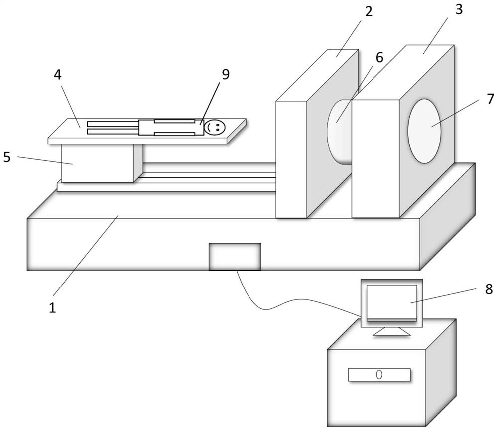
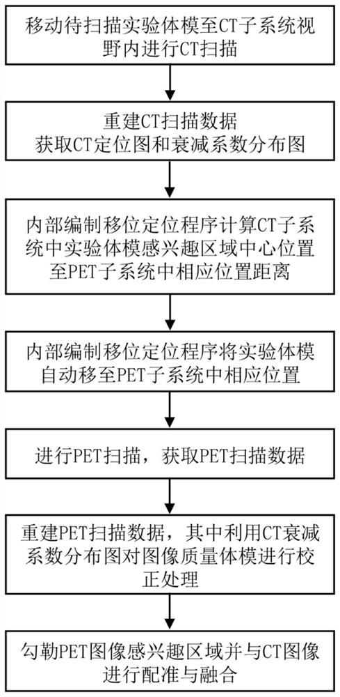
[0044] When the experimental object 9 to be scanned of the present invention is a phantom, the following steps can be performed according to the attributes of the experimental object:

[0045] ① Operate the scan control and image processing workstation 8 to move the phantom to the CT subsystem 3 to complete the CT scan and obtain CT scan data;

[0046] ② Reconstruct the CT scan data to obtain the corresponding CT positioning image and attenuation coefficient distribution map;

[0047] ③According to the CT positioning image obtained in step ②, use the displacement positioning program compiled in the dual-mode PET / CT system to calculate the distance between the center position of the region of interest in the phantom of the CT subsystem and the corresponding position in the PET scanning part. the distance L;

[0048] ④According to the distance L obtained in step ③, use the displacement positioning program compiled in the dual-mode PET / CT system to automatically move the area of...

Embodiment 2

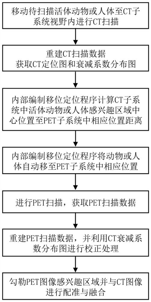
[0058] When the subject 9 to be scanned of the present invention is a living animal or a human body, the following steps can be performed according to the properties of the subject:

[0059] ① Operate the scan control and image processing workstation 8 to move the live animal or human body to the CT subsystem 3 to complete the CT scan and obtain the CT scan data;

[0060] ② Reconstruct the CT scan data to obtain the corresponding CT positioning image and attenuation coefficient distribution map;

[0061] ③According to the CT positioning image obtained in step ②, use the displacement positioning program compiled in the dual-mode PET / CT system to calculate the difference between the center position of the region of interest in the CT scanning part of the living animal or human body and the corresponding position in the PET scanning part. the distance L between

[0062] ④According to the distance L obtained in step ③, use the displacement positioning program compiled in the dual...


PUM


Abstract

The invention discloses an automatic precise positioning scanning method of a PET / CT system. The automatic precise positioning scanning method comprises the following steps: moving an experimental object to a CT subsystem to complete CT scanning, and obtaining CT scanning data; reconstructing CT scanning data to obtain a CT positioning image and an attenuation coefficient distribution diagram; according to the obtained CT positioning image, calculating the distance L between the central position of the region of interest of the experimental subject in the CT scanning part and the corresponding position in the PET scanning part by using a displacement positioning program of the PET / CT system; using the PET / CT system for automatically moving the region of interest of the experimental subject to a PET subsystem part for positioning; conducting PET scanning, and obtaining a corresponding PET image; and drawing a region of interest in the PET image and conducting registration and fusion with the CT image. According to the automatic precise positioning scanning method, automatic accurate positioning is realized, radiation to personnel is reduced, specific scanning imaging is conducted on a local region of interest, bed scanning data image reconstruction time is shortened, and scanning imaging progress of a scanned object is accelerated.

Description

technical field [0001] The invention relates to a PET / CT system, in particular to a PET / CT system automatic and precise positioning scanning method, which belongs to the technical field of medical imaging equipment. Background technique [0002] PET / CT, a dual-mode medical imaging device combining Positron Emission Tomography (PET) and Computed Tomography (CT), is currently the most important tool for preclinical research and clinical practice to diagnose and guide treatment. advanced means. The image information obtained by CT imaging of anatomical structures can provide clinical information on the internal anatomical structure of the patient's physiological form, generally has a high image resolution (below 1mm), but lacks functional metabolic information, and cannot diagnose early tissue lesions and subtle lesions ; The image information collected by functional metabolic PET imaging can provide clinical information on the patient's metabolic function and perform accurate...

Claims


Application Information

Patent Timeline
Patent Type & Authority Applications(China)
IPC IPC(8): A61B6/03A61B6/04
CPCA61B6/032A61B6/037A61B6/0407A61B6/54A61B6/545
Inventor 陈志成刘琼李超凡王德广
Owner XUZHOU MEDICAL UNIV
Learn More