Lymph node metastasis image analysis system, method and equipment based on deep learning

A technology of lymph node metastasis and deep learning, applied in neural learning methods, image analysis, image enhancement, etc., can solve problems such as non-invasive prediction of non-small cell carcinoma images, achieve good robustness and generalization ability, and improve prognosis. Effect

Active Publication Date: 2021-06-18
INST OF AUTOMATION CHINESE ACAD OF SCI
View PDF7 Cites 4 Cited by
  • Summary
  • Abstract
  • Description
  • Claims
  • Application Information

AI Technical Summary

Problems solved by technology

[0007] In order to solve the above-mentioned problems in the prior art, that is, the prior art cannot perform non-invasive prediction of whether lymph node metastasis occurs in non-small cell carcinoma images, the present invention extracts the lesion and its microenvironment area, and Integrate CT image data, patient clinical indicators and

Method used


Image

  • Lymph node metastasis image analysis system, method and equipment based on deep learning
  • Lymph node metastasis image analysis system, method and equipment based on deep learning
  • Lymph node metastasis image analysis system, method and equipment based on deep learning

Examples


Embodiment Construction

[0062] The application will be further described in detail below in conjunction with the accompanying drawings and embodiments. It should be understood that the specific embodiments described here are only used to explain related inventions, not to limit the invention. It should also be noted that, for the convenience of description, only the parts related to the related invention are shown in the drawings.

[0063] It should be noted that, in the case of no conflict, the embodiments in the present application and the features in the embodiments can be combined with each other. The present application will be described in detail below with reference to the accompanying drawings and embodiments.

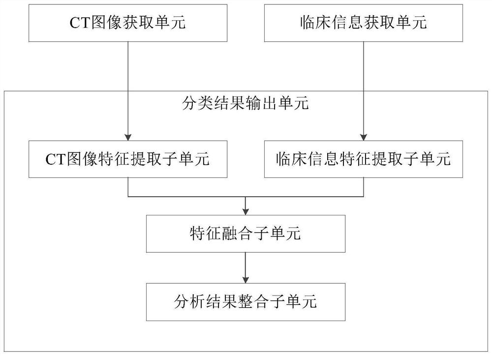
[0064] The present invention provides an image analysis system for lymph node metastasis based on deep learning. The system includes: a CT image acquisition unit, a clinical information acquisition unit and a classification result output unit;

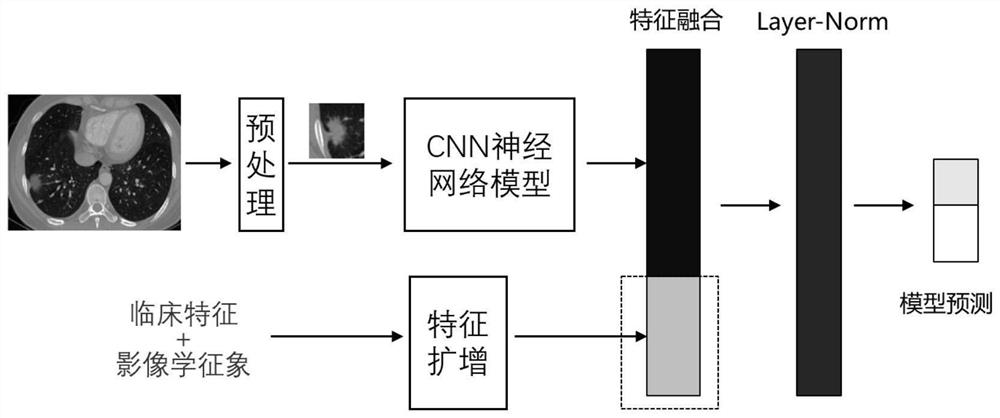
[0065] The CT image acquisition unit ...


PUM


Abstract

The invention belongs to the field of image analysis, particularly relates to a lymph node metastasis image analysis system, method and equipment based on deep learning, and aims to solve the problem that in the prior art, whether lymph node metastasis occurs in a non-small cell carcinoma image cannot be well predicted noninvasively. The method comprises the steps of obtaining a to-be-analyzed CT image containing a lesion microenvironment, obtaining an imaging sign and clinical information of the same subject as the CT image, extracting one-dimensional CT image features and one-dimensional clinical information features respectively, performing feature enhancement and normalization processing, performing fusion through a full connection layer to generate a fusion feature vector, and classifying the fused feature vectors to obtain an analysis result. The non-small cell lung cancer lymph node metastasis image data classification method achieves classification of non-small cell lung cancer lymph node metastasis image data, has good robustness and generalization ability compared with an existing traditional vector model based on predefined image features, and effectively improves the prognosis effect of a patient.

Description

technical field [0001] The invention belongs to the field of image analysis, and in particular relates to a deep learning-based lymph node metastasis image analysis system, method and equipment. Background technique [0002] Lung cancer is one of the malignant tumors with the fastest-growing morbidity and mortality. Due to differences in gender, age, race, and geographical location, the incidence of lung cancer also varies greatly. Its main subtypes include squamous cell carcinoma and large cell carcinoma. and lung adenocarcinoma. [0003] For patients with early-diagnosed non-small cell lung cancer, if a more accurate preoperative classification can be obtained, the postoperative survival period is usually longer and the quality of life is also higher. Especially for the lymph node metastasis of early non-small cell lung cancer, it is difficult to find the effective characteristics of lymph node metastasis by naked eyes through CT and other imaging methods. Therefore, the ...

Claims


Application Information

Patent Timeline
IPC IPC(8): G06T7/00G06T7/11G06K9/46G06K9/62G06N3/08
CPCG06T7/0012G06T7/11G06N3/08G06T2207/10081G06T2207/30061G06T2207/30096G06T2207/20081G06V10/40G06F18/24G06F18/253Y02A90/10
Inventor 田捷董迪李海林胡振华王思雯胡朝恩
Owner INST OF AUTOMATION CHINESE ACAD OF SCI
Why Eureka
Try Eureka