Cell smear auxiliary analysis method and system

A technology for auxiliary analysis and cell smear, applied in the field of digital image processing technology and computer-aided medical examination, it can solve the problems of difficult localization and traceability, only based on operation memory, deviation, etc. The effect of running speed and reducing the amount of calculation

Active Publication Date: 2021-08-13
SOUTH CHINA NORMAL UNIVERSITY
View PDF13 Cites 0 Cited by
  • Summary
  • Abstract
  • Description
  • Claims
  • Application Information

AI Technical Summary

Problems solved by technology

[0003] In this method, the professional quality of inspectors is high, the process is cumbersome and time-consuming, and the analysis results are easily affected by subjective factors. Since the process of operating a microscope to obtain a series of cell images is only restricted by simple moving direction rules, the operation in this process is controlled by the inspector. At the same time, the movement of the microscope stage has a position offset in the direction perpendicular to the movement direction due to the mechanical structure, and in the traditional microscope operation, the inspection observation cannot be recorded because the stage movement is not recorded. In the process of operation, the inspector judges the position of the field of view under the microscope only according to the operation memory, which has a large deviation, and it is difficult to locate the previously inspected area or cell during or after the inspection and traceability
[0004] At the same time, if there are doubts about the judgment of a certain cell in the cell smear, when multiple inspectors are required to verify, it is necessary to conduct a second observation of the area where the cell is located. In the traditional method, it is difficult to verify the cell after changing the inspector or changing the microscope. Precise positioning and field of view backtracking in the area where the cells are located

Method used


Image

  • Cell smear auxiliary analysis method and system
  • Cell smear auxiliary analysis method and system
  • Cell smear auxiliary analysis method and system

Examples


Embodiment 1

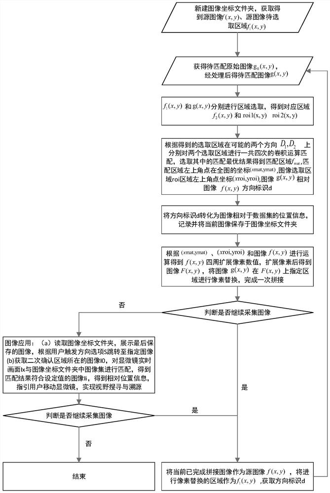
[0196] Such as figure 1 As shown, the present invention provides a cell smear auxiliary analysis method, comprising the steps of:

[0197] Step S1: Create a folder and initialize parameters to obtain the first cell image;

[0198] S11: Create a new folder named image coordinate folder, which is used to store the cell image set I containing the position information loc(a,b) obtained during the operation of the system;

[0199] S12: Record the moving direction of the microscope field of view selected by the user as a direction mark d, and according to the following scanning rules, the direction mark d is one of "right", "left" and "down";

[0200] S13: Taking the first cell image through the microscope I 1 , select as the source image f(x,y), assign the position information loc(a,b) to the image as loc(0,0) and save it in the image coordinate folder;

[0201] S13: Use the whole image of the source image f(x,y) as the area f to be selected in the source image 1 (x,y);

[020...

Embodiment 2

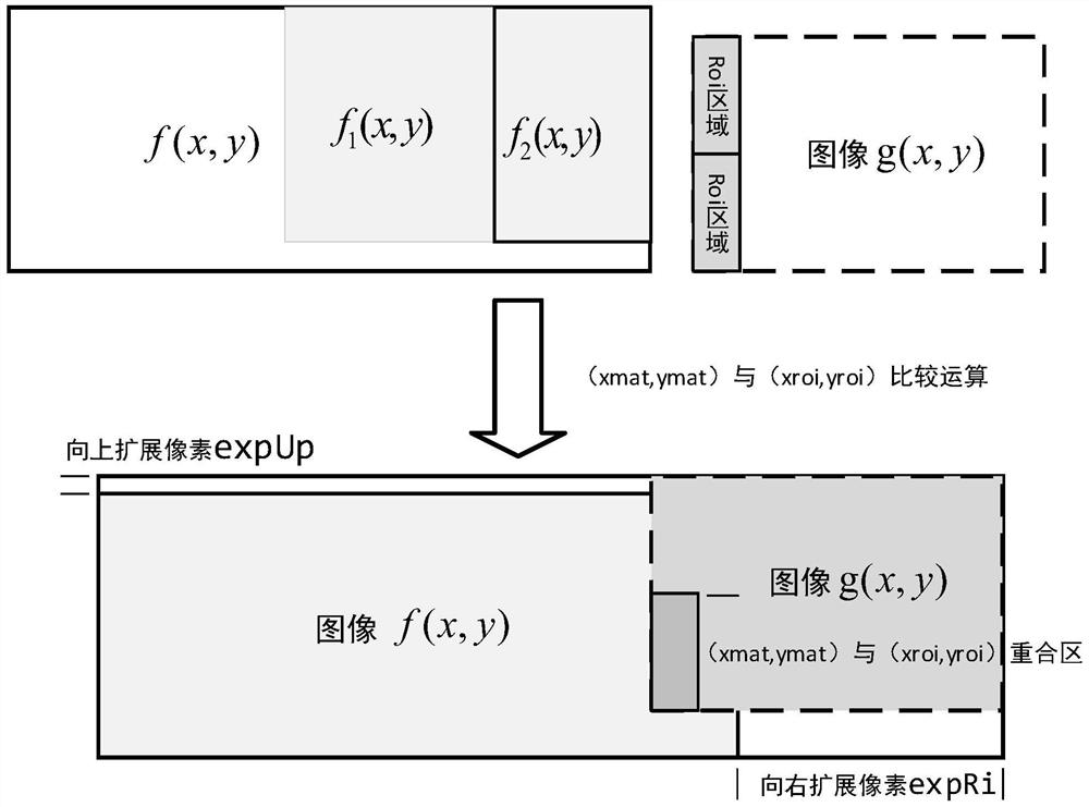
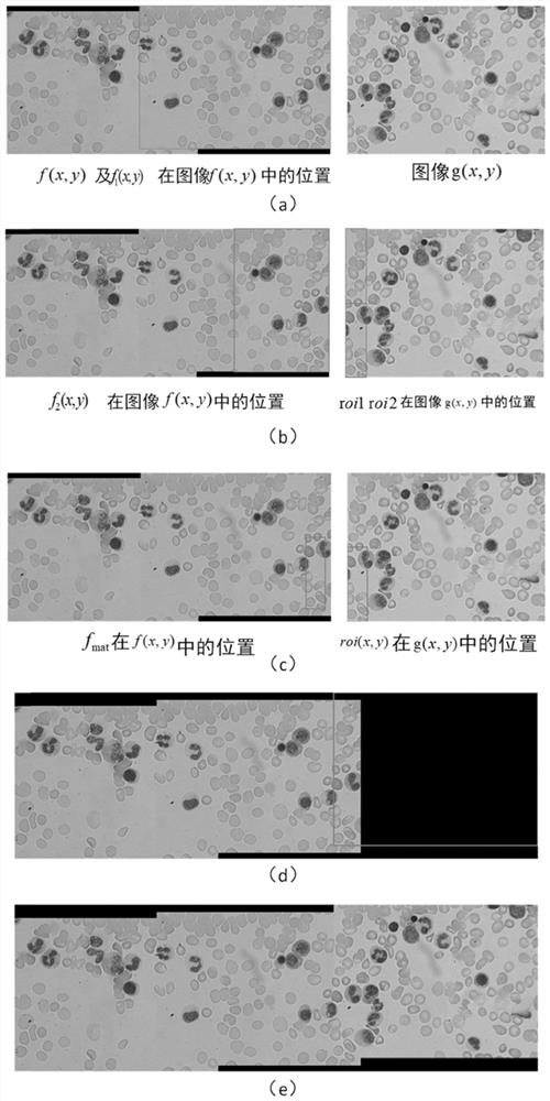
[0281] Such as Figure 5 As shown, the present invention provides a cell smear auxiliary analysis system, including an image input module, an area matching module, an image stitching module and an image application module;

[0282] The image input module is externally connected to a microscope, which is used for continuous collection of cell images by the microscope camera, and a cell image is taken through the microscope, selected as the source image, and the entire image of the source image is used as the area to be selected in the source image; press "From left to Right, moving the microscope regularly in a serpentine shape from top to bottom to acquire a cell image that has an m% overlapping area with the field of view of the previous image, select it as the original image to be matched, and preprocess the original image to be matched to obtain the image to be matched;

[0283] The region matching module is used to identify the possible splicing direction through the direc...


PUM


Abstract

The invention discloses a cell smear auxiliary analysis method and system. The method comprises the following steps: continuously collecting cell images by a microscope camera; identifying a possible splicing direction through a direction identifier, performing region selection and matching on the two images participating in splicing, and obtaining a matching result through calculation; carrying out image pixel expansion according to a matching result, and carrying out pixel replacement in a matching area after expansion; converting image relative position information obtained in the splicing process into numerical information recorded in a coordinate mode, and constructing a coordinate system of the images in the image set; displaying the spliced image to a user, wherein the user can visually see the condition of the currently observed cell region; and at the same time, according to the obtained image coordinate system, providing re-observation of the checked cell image area, so that when the current image is displayed, the image area around the visual field in the corresponding checking process is jumped to according to the direction option triggered by the user. The method and system convenient to use, high in interactivity and high in operation speed.

Description

technical field [0001] The invention relates to the technical fields of digital image processing technology and computer-aided medical examination, in particular to a cell smear-assisted analysis method and system. Background technique [0002] Cytomorphological examination is an important way for the diagnosis of blood diseases. Statistical analysis of the morphology, type and quantity of blood cell smears plays an important role in the diagnosis of various diseases. The traditional method of analyzing and counting the number of cells in a smear is: the inspector makes a slide smear from the patient's peripheral blood or bone marrow blood, stains it and places it on the microscope stage, and observes the number of cells in the field of view of the microscope eyepiece. Morphology, analyze its type and record the number, then move the smear according to the designated moving direction, record the type and number of cells in the current field of view, and repeat this process u...

Claims


Application Information

Patent Timeline
IPC IPC(8): G06T7/73G01N15/10
CPCG06T7/74G01N15/10G06T2207/10056G06T2207/30024G01N2015/1006G01N2015/1022G01N2015/1024
Inventor 马琼雄李梓岚何海森苏志豪陆清航张庆茂
Owner SOUTH CHINA NORMAL UNIVERSITY
Why Eureka
Try Eureka