A sort of 68 Ga-labeled molecular probe, preparation method and application thereof
A molecular probe, 68ga technology, applied in the field of nuclear medicine imaging, achieves good stability, good application prospects, and excellent imaging effects
- Summary
- Abstract
- Description
- Claims
- Application Information
AI Technical Summary
Problems solved by technology
Method used
Image
Examples
Embodiment 1
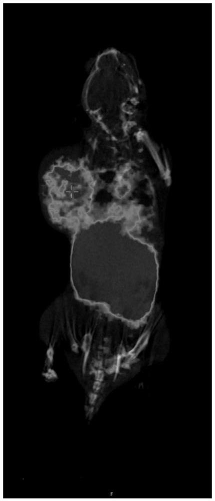
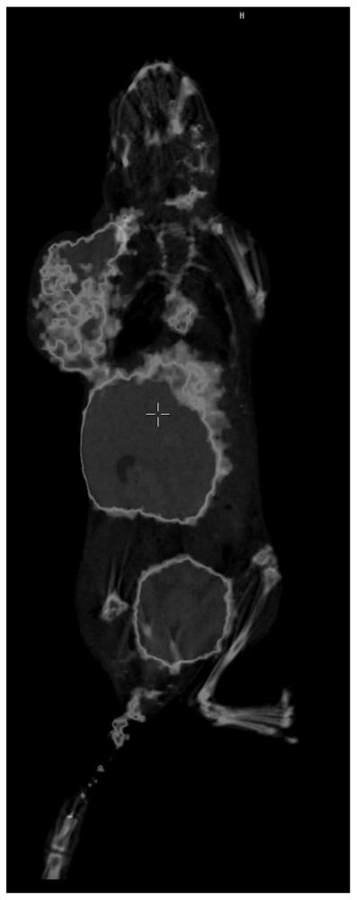
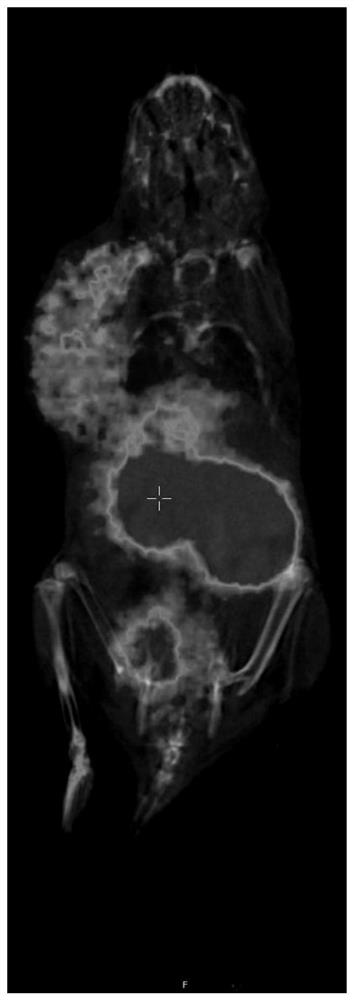
[0032] The preparation method of the molecular imaging agent described in this embodiment is as follows:
[0033] 1) Put 25ug of the precursor compound NOTA-Spermin into the EP tube, add 1ml of 0.25M NaOAc solution, mix well and transfer to the reaction tube;
[0034] 2) with 4ml of 0.05M HCl solution 68 GaCl3 is rinsed into the reaction tube, after rinsing 68 The Ga radioactivity was 31mCi, and the reaction was carried out at 90 °C for 10 min. After the reaction, 10 ml of deionized water was added to quench the reaction;
[0035] 3) The solution in the reaction tube is enriched through the C18 Plus column, and the C18 Plus column is washed with 10 mL of deionized water; then the C18 Plus column is washed with 1 ml of ethanol and 10 ml of normal saline to obtain an eluent, and the eluent is passed through without After bacterial filtration membrane, it is loaded into the product bottle to obtain the imaging agent injection 68 Ga-NOTA-Spermin.
[0036] Imaging agent injecti...
Embodiment 2
[0041] The preparation method of the molecular imaging agent described in this embodiment is as follows:
[0042] 1) Put 30ug of the precursor compound NOTA-Spermidine into the EP tube, add 1ml of 0.25M NaOAc solution, mix well and transfer it to the reaction tube;
[0043] 2) with 4ml of 0.05M HCl solution 68 GaCl3 is rinsed into the reaction tube, after rinsing 68 The Ga radioactivity was 33mCi, and the reaction was carried out at 90 °C for 10 min. After the reaction, 10 ml of deionized water was added to quench the reaction;
[0044] 3) The solution in the reaction tube is enriched through the C18 Plus column, and the C18 Plus column is washed with 10 mL of deionized water; then the C18 Plus column is washed with 1 ml of ethanol and 10 ml of normal saline to obtain an eluent, and the eluent is passed through without After bacterial filtration membrane, it is loaded into the product bottle to obtain the imaging agent injection 68 Ga-NOTA-Spermidine.
[0045] Imaging agen...
Embodiment 3
[0050] The preparation method of the molecular imaging agent described in this embodiment is as follows:
[0051] 1) Put 30ug of the precursor compound NOTA-Putrescine into an EP tube, add 1ml of 0.25M NaOAc solution, mix well and transfer it to a reaction tube;
[0052] 2) with 4ml of 0.05M HCl solution 68 GaCl3 is rinsed into the reaction tube, after rinsing 68 The Ga radioactivity was 35mCi, and the reaction was carried out at 90 °C for 10 min. After the reaction, 10 ml of deionized water was added to quench the reaction;
[0053] 3) The solution in the reaction tube is enriched through the C18 Plus column, and the C18 Plus column is washed with 10 mL of deionized water; then the C18 Plus column is washed with 1 ml of ethanol and 10 ml of normal saline in turn to obtain an eluent, and the eluent is passed through without After bacterial filtration membrane, it is loaded into the product bottle to obtain the imaging agent injection 68 Ga- NOTA-Putrescine.
[0054] Imagin...
PUM
| Property | Measurement | Unit |
|---|---|---|
| radioactivity | aaaaa | aaaaa |
| radioactivity | aaaaa | aaaaa |
| radioactivity | aaaaa | aaaaa |
Abstract
Description
Claims
Application Information
 - R&D
- Intellectual Property
- Life Sciences
- Materials
- Tech Scout
- Unparalleled Data Quality
- Higher Quality Content
- 60% Fewer Hallucinations
Browse by: Latest US Patents, China's latest patents, Technical Efficacy Thesaurus, Application Domain, Technology Topic, Popular Technical Reports.
© 2025 PatSnap. All rights reserved.Legal|Privacy policy|Modern Slavery Act Transparency Statement|Sitemap|About US| Contact US: help@patsnap.com


