Postoperative tumor evaluation method, device and computer storage medium

A tumor and imaging technology, applied in computer components, computer-aided medical procedures, calculations, etc., can solve problems such as delaying supplementary treatment time for high-risk patients, affecting long-term estimation, and being unable to predict tumor recurrence risk

Active Publication Date: 2021-09-10
MAGI CO LTD
View PDF12 Cites 0 Cited by
  • Summary
  • Abstract
  • Description
  • Claims
  • Application Information

AI Technical Summary

Problems solved by technology

However, manual diagnosis based on imaging follow-up can only measure the changes that have occurred during imaging and can reflect macroscopic tissue changes, ignoring the potential manifestations of tumor microcirculation damage and antigen release in imaging, which affects the long-term estimation of prognosis
[0008] Third, the current prognosis assessment method of tumor ablation is difficult to immediately evaluate whether the operation is successful after the end of treatment, nor can it predict the risk of tumor recurrence over time. Guidance reference, which will delay the optimal time for supplementary treatment in high-risk patients, bringing potential risk of relapse

Method used


Image

  • Postoperative tumor evaluation method, device and computer storage medium
  • Postoperative tumor evaluation method, device and computer storage medium
  • Postoperative tumor evaluation method, device and computer storage medium

Examples


no. 1 example

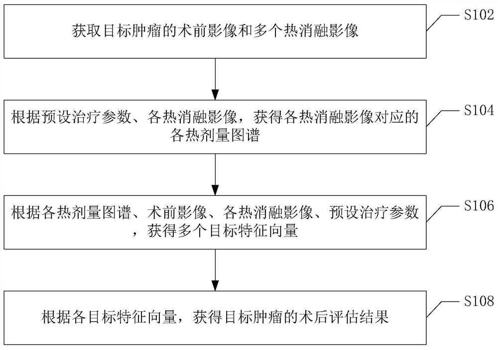
[0029] figure 1 It is a schematic flowchart of the postoperative tumor assessment method according to the first embodiment of the present application. As shown in the figure, the postoperative tumor method in this embodiment mainly includes:

[0030] Step S102, acquiring a preoperative image and a plurality of thermal ablation images of the target tumor.

[0031] Optionally, a preoperative image of the target tumor before ablation is performed, and thermal ablation images of the target tumor corresponding to each thermal ablation operation are acquired.

[0032] In this embodiment, the ablation performed on the target tumor may include ablation treatment schemes such as cold ablation alone, thermal ablation alone, and a combination of cold ablation and thermal ablation.

[0033] Wherein, each thermal ablation operation (such as radio frequency ablation) corresponds to different ablation positions of the target tumor respectively.

[0034] Optionally, a cold ablation image o...

no. 2 example

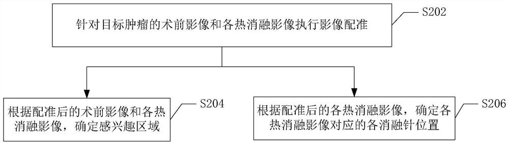
[0052] figure 2 A schematic flowchart of the second embodiment of the present application is shown. As shown in the figure, the postoperative tumor assessment method of this embodiment can be performed after step S102 and before step S104, which mainly includes the following steps:

[0053] Step S202, image registration is performed on the preoperative image of the target tumor and each thermal ablation image.

[0054] In this embodiment, based on the thermal ablation image of the first thermal ablation operation, deformable registration can be performed on the preoperative image, and rigid registration can be performed on other thermal ablation images.

[0055] Specifically, according to the imaging difference of the same tissue to be ablated (target tumor) in the image sequence, an appropriate image registration method can be selected to correct body position changes and multiple needle advance and retreat operations during treatment (one thermal ablation operation will ge...

no. 3 example

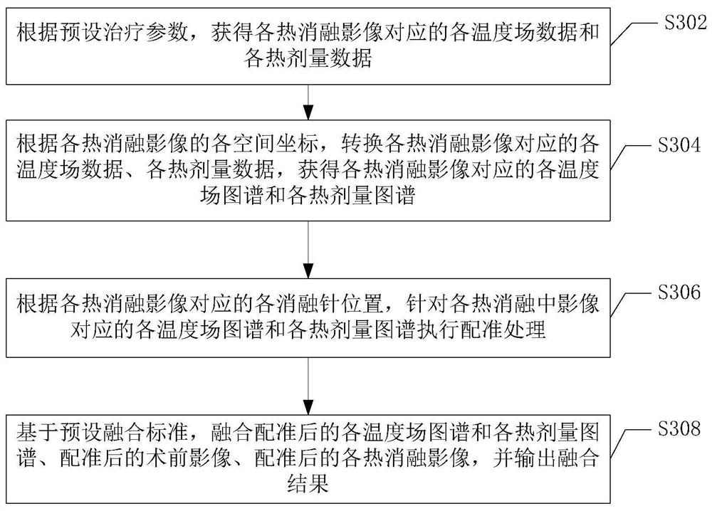
[0068] image 3 A schematic flowchart of the postoperative tumor assessment method according to the third embodiment of the present application is shown. This embodiment is a specific implementation of the above step S104, which mainly includes the following steps:

[0069] In step S302, according to preset treatment parameters, each temperature field data and each thermal dose data corresponding to each thermal ablation image are obtained.

[0070] Optionally, the preset simulation model can be configured according to the preset treatment parameters, and based on the configured preset simulation model, each temperature field data and each thermal dose corresponding to each voxel block in the region of interest of the thermal ablation image can be obtained data.

[0071] In this embodiment, the preset simulation model can be used to invert the accumulated steady-state temperature field and thermal dose distribution from the beginning of each thermal ablation operation accord...


PUM


Abstract

The invention provides a postoperative tumor evaluation method, a device and a computer storage medium. The postoperative tumor evaluation method mainly comprises the steps of obtaining a preoperative image and a plurality of thermal ablation images of a target tumor; according to preset treatment parameters and the thermal ablation images, obtaining thermal dose maps corresponding to the thermal ablation images; obtaining a plurality of target feature vectors according to the thermal dose maps, the preoperative image, the thermal ablation images and the preset treatment parameters; and obtaining a postoperative evaluation result of the target tumor according to each target feature vector. Therefore, the postoperative tumor recurrence risk can be quickly and accurately predicted, so that a clinician can be assisted to more accurately master the condition of a patient.

Description

technical field [0001] The embodiments of the present application relate to the technical field of artificial intelligence, and in particular to a postoperative tumor assessment method, device, and computer storage medium. Background technique [0002] Tumor ablation is a minimally invasive treatment technique with high safety and few complications. It is an important option for treating cancer patients who have failed chemotherapy and radiotherapy or are not suitable for surgical resection. [0003] Existing ablation equipment can directly transmit energy to the target tissue and cause physical damage in situ: among them, thermal ablation therapy refers to heating the target tissue to a temperature higher than 60°C, causing rapid denaturation of proteins and cell nuclei; cryoablation therapy is based on the Joule-Thomson principle, cooling the target tissue to below -40°C to destroy the cell structure and cause apoptosis and necrosis; in addition, combined heating and cooli...

Claims


Application Information

Patent Timeline
Patent Type & Authority Applications(China)
IPC IPC(8): G06K9/62G06K9/46G16H50/30
CPCG16H50/30G06F18/2411Y02A90/10
Inventor 不公告发明人
Owner MAGI CO LTD
Why Eureka
Try Eureka