Eureka AIR delivers breakthrough ideas for toughest innovation challenges, trusted by R&D personnel around the world.

Aorta structure image automatic segmentation method based on artificial intelligence

A structural image and automatic segmentation technology, applied in the field of medical image processing, can solve the problems of inaccurate measurement, difficult to reproduce and reproduce, and measurement subjectivity, etc., to improve efficiency and accuracy, improve the perfect and accurate segmentation of incompletely segmented areas Effect

Active Publication Date: 2021-09-17
拓微摹心数据科技(北京)有限公司
View PDF7 Cites 1 Cited by
  • Summary
  • Abstract
  • Description
  • Claims
  • Application Information

AI Technical Summary

Problems solved by technology

It solves the shortcomings of inaccurate manual measurement, high subjectivity of measurement, human error, and difficulty in copying and reproducing.

Method used


Image

  • Aorta structure image automatic segmentation method based on artificial intelligence
  • Aorta structure image automatic segmentation method based on artificial intelligence
  • Aorta structure image automatic segmentation method based on artificial intelligence

Examples


Embodiment Construction

[0058] Below in conjunction with specific embodiment, further illustrate the present invention. It should be understood that the examples are only used to illustrate the present invention and not to limit the protection scope of the present invention. In addition, it should be understood that after reading the disclosure of the present invention, those skilled in the art may make various changes or modifications to the present invention, and these equivalent forms also fall within the scope of protection defined by the present invention.

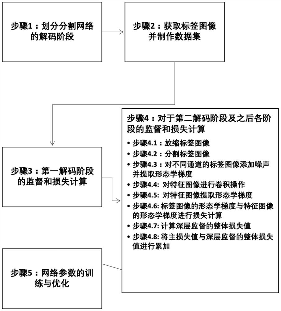
[0059] Such as figure 1 As shown, the artificial intelligence-based aortic structure image automatic segmentation method of the present invention comprises the following 5 steps:

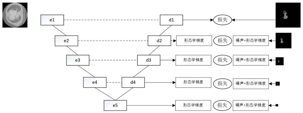
[0060] Step 1: Divide the decoding stage of the segmentation network, specifically divided into 4 or 5 stages, adopt 5 decoding stages in this embodiment, such as figure 2 shown.

[0061] Step 2: Obtain the labeled image and make a data set. According to the o...


PUM


Abstract

The invention discloses an aorta structure image automatic segmentation method based on artificial intelligence. The method the steps of segmenting label images, adding noise to the label images of different channels and extracting morphological gradients, performing morphological feature extraction on multi-channel feature images of each decoding stage, performing loss calculation and total loss calculation of each stage, and finally completing training and optimization of network parameters. Compared with the existing image processing method, the image processing method based on artificial intelligence has the advantages that obvious mistakenly segmented areas can be removed, the image segmentation effect can be improved, the target area can be segmented more accurately, the incompletely segmented area is more perfect. Image data with higher accuracy is provided for later establishment of a three-dimensional model, and the efficiency and precision of TAVR / TAVI preoperative evaluation are effectively improved.

Description

technical field [0001] The invention belongs to the field of medical image processing, and in particular relates to an artificial intelligence-based automatic segmentation method for aortic images, which is mainly applied to preoperative evaluation of transcatheter aortic valve replacement. Background technique [0002] The aortic root is located in the center of the heart, below the aortic sinus. The aortic sinus is inserted between the mitral valve and the tricuspid valve in a cylindrical shape, the base is completely embedded in the surrounding tissue, and the second half of the circle is completely surrounded by the two atria. The coronary arteries that supply blood to the heart itself generally open into the left and right coronary sinuses within the aortic sinus. The aortic valve is located at the bottom of the aortic root, at the junction of the aortic sinus and the left ventricular outflow tract, which constitutes the boundary between the aorta and the left ventricl...

Claims


Application Information

Patent Timeline
IPC IPC(8): G06T7/155G06N3/04G06N3/08G06T5/00G06T7/11G06T9/00
CPCG06T7/155G06T7/11G06N3/08G06T9/00G06T2207/10081G06T2207/30101G06T2207/30048G06N3/045G06T5/70
Inventor 马琛明方桧铭邓智方
Owner 拓微摹心数据科技(北京)有限公司
Why Eureka
Eureka Blog
Learn More