Eureka AIR delivers breakthrough ideas for toughest innovation challenges, trusted by R&D personnel around the world.

Craniocerebral CT image bleeding area automatic segmentation method

An automatic segmentation and CT image technology, applied in the field of medical image processing, can solve the problems of low accuracy and slow image recognition speed

Pending Publication Date: 2020-09-29
SHANGHAI INST OF TECH
View PDF5 Cites 4 Cited by
  • Summary
  • Abstract
  • Description
  • Claims
  • Application Information

AI Technical Summary

Problems solved by technology

[0004] The technical purpose of the present invention is to provide a method for automatic segmentation of hemorrhage areas in cranial CT images to solve the technical problems of slow image recognition speed and low precision

Method used


Image

  • Craniocerebral CT image bleeding area automatic segmentation method
  • Craniocerebral CT image bleeding area automatic segmentation method
  • Craniocerebral CT image bleeding area automatic segmentation method

Examples


Embodiment Construction

[0040] In order to more clearly illustrate the embodiments of the present invention or the technical solutions in the prior art, the specific implementation manners of the present invention will be described below with reference to the accompanying drawings. Obviously, the accompanying drawings in the following description are only some embodiments of the present invention, and those skilled in the art can obtain other accompanying drawings based on these drawings and obtain other implementations.

[0041] In order to make the drawing concise, each drawing only schematically shows the parts related to the present invention, and they do not represent the actual structure of the product. In addition, to make the drawings concise and easy to understand, in some drawings, only one of the components having the same structure or function is schematically shown, or only one of them is marked. Herein, "a" not only means "only one", but also means "more than one".

[0042] A method f...


PUM


Abstract

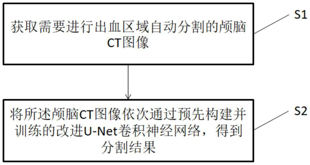
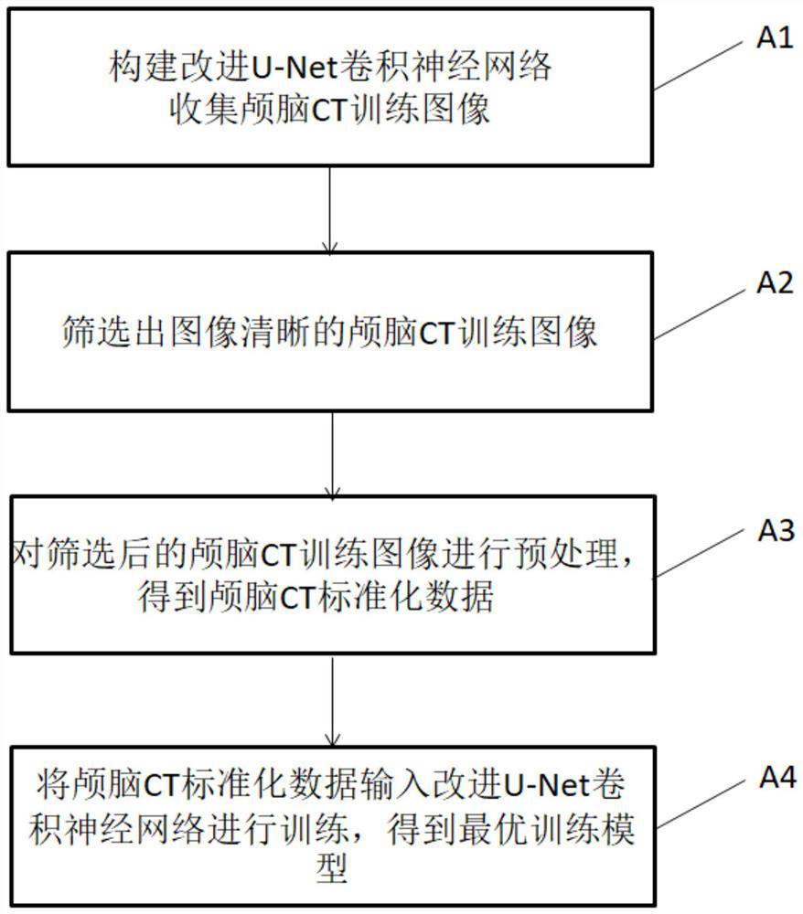
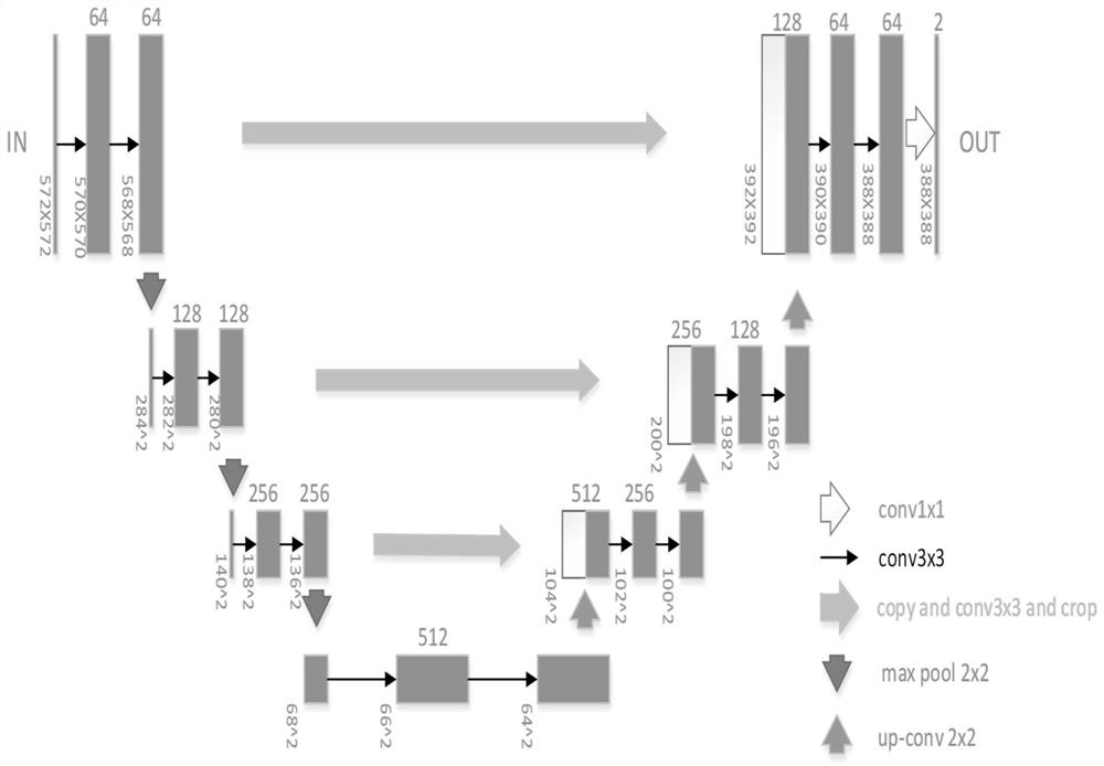
The invention discloses a craniocerebral CT image bleeding area automatic segmentation method. The method comprises the steps of: S1, obtaining a craniocerebral CT image needing bleeding area automatic segmentation; and S2, enabling the craniocerebral CT image to sequentially pass through a pre-constructed and trained improved U-Net convolutional neural network, wherein the overall structure of the network is three down-sampling layers and three up-sampling layers, and in the jump connection process, feature maps obtained through down-sampling are copied, convolved and cut respectively and then spliced with feature maps behind the corresponding up-sampling layers, and the corresponding up-sampling layers carry out up-sampling and convolution on the spliced feature maps. The improved U-Netconvolutional neural network totally comprises seven layers, so that information loss is reduced while feature extraction is ensured, time is saved, and the overall efficiency is improved; and in thejump connection step, convolution operation is added to a down-sampled feature map, more information can be provided for an up-sampling layer while the number of model layers is not changed, and therefore subsequent image segmentation of the cerebral hemorrhage CT image is improved.

Description

technical field [0001] The invention belongs to the field of medical image processing, and in particular relates to a method for automatically segmenting hemorrhage areas in cranial CT images. Background technique [0002] According to the Global Burden of Disease, stroke is the second most common cause of death and one of the leading causes of disability worldwide. A stroke occurs when blood flow to a specific area of ​​the brain is interrupted. When this happens, brain cells are deprived of oxygen and die. When a stroke causes brain cells to die, functions controlled by this area of ​​the brain, such as memory and muscle control, are lost. A hemorrhagic stroke occurs when a blood vessel ruptures, while an ischemic stroke occurs when a blood vessel becomes blocked, causing more damage than an ischemic stroke. [0003] In the medical field, in order to meet the needs of disease diagnosis and treatment plan formulation, it is often necessary to scan patients to determine th...

Claims


Application Information

Patent Timeline
Patent Type & Authority Applications(China)
IPC IPC(8): G06T7/11
CPCG06T7/11G06T2207/10081G06T2207/20081G06T2207/20084G06T2207/30016
Inventor 曹国刚王一杰朱信玉李梦雪曹聪刘顺堃毛红东孔德卿
Owner SHANGHAI INST OF TECH
Why Eureka
Eureka Blog
Learn More