
Brain tumor detection device based on magnetic resonance electrical impedance tomography

An electrical impedance imaging, brain tumor technology, applied in the medical field, can solve the problems of non-standard rotation, reduced detection effect, no detection device cleaning and disinfection, etc., to achieve the effect of improving the use effect and improving the accuracy.

Pending Publication Date: 2021-10-01
管晓翔
View PDF6 Cites 0 Cited by
  • Summary
  • Abstract
  • Description
  • Claims
  • Application Information

AI Technical Summary

Problems solved by technology

[0003] The existing brain tumor detection device based on magnetic resonance electrical impedance imaging does not have the function of cleaning and disinfecting the detection device, which may cause the generation of germs during use and reduce the use effect of the detection device. , the patient needs to turn the body by himself, and when the patient turns by himself, the rotation may not be standard, which reduces the detection effect

Method used


Image

  • Brain tumor detection device based on magnetic resonance electrical impedance tomography
  • Brain tumor detection device based on magnetic resonance electrical impedance tomography
  • Brain tumor detection device based on magnetic resonance electrical impedance tomography

Examples


Embodiment 1

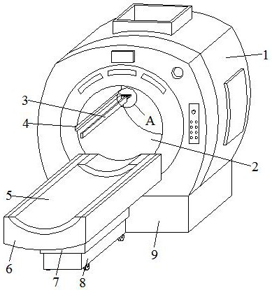
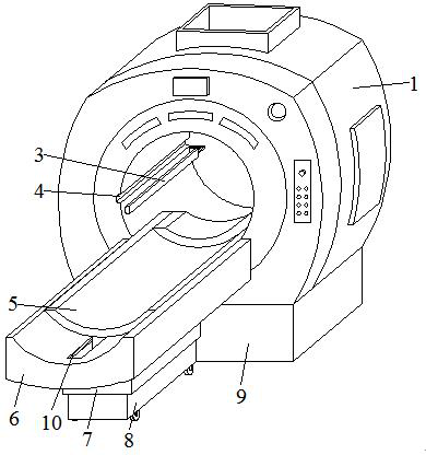
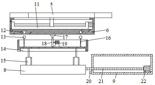
[0030] refer to Figure 1-7 , a brain tumor detection device based on magnetic resonance electrical impedance imaging, comprising a main body 1, a base 9 and a strip plate 8, a through hole 2 is opened on one side of the main body 1, and the base 9 is horizontally and fixedly connected to the bottom of the main body 1 , the strip plate 8 is located on one side of the base, and the inside of the base 9 is provided with a horizontal movement mechanism for horizontally moving the strip plate 8, the inner wall of the through hole 2 is horizontally provided with a strip groove 4, and the inside of the strip groove 4 slides. For the matching brush plate 3, a first rotating mechanism for rotating the brush plate 3 is provided inside the main body 1, and a hollow box 7 is horizontally arranged directly above the strip plate 8, and a rectangular groove 14 is opened at the bottom of the hollow box 7, and Both ends of the top of the strip plate 8 are provided with a lifting mechanism for...

Embodiment 2

[0034] refer to Figure 8 , a brain tumor detection device based on magnetic resonance electrical impedance imaging, comprising a main body 1, a base 9 and a strip plate 8, a through hole 2 is opened on one side of the main body 1, and the base 9 is horizontally and fixedly connected to the bottom of the main body 1 , the strip plate 8 is located on one side of the base, and the inside of the base 9 is provided with a horizontal movement mechanism for horizontally moving the strip plate 8, the inner wall of the through hole 2 is horizontally provided with a strip groove 4, and the inside of the strip groove 4 slides. For the matching brush plate 3, a first rotating mechanism for rotating the brush plate 3 is provided inside the main body 1, and a hollow box 7 is horizontally arranged directly above the strip plate 8, and a rectangular groove 14 is opened at the bottom of the hollow box 7, and Both ends of the top of the strip plate 8 are provided with a lifting mechanism for l...


PUM


Abstract

The invention discloses a brain tumor detection device based on magnetic resonance electrical impedance tomography. The brain tumor detection device comprises a main body, a base and a strip-shaped plate, a through hole is formed in one side of the main body, the base is horizontally and fixedly connected to the bottom of the main body, the strip-shaped plate is located on one side of the base, and a horizontal moving mechanism used for horizontally moving the strip-shaped plate is arranged in the base; a strip-shaped groove is horizontally formed in the inner wall of the through hole, a brush plate matched with the strip-shaped groove is slidably arranged in the strip-shaped groove, a first rotating mechanism used for rotating the brush plate is arranged in the main body, a hollow box is horizontally arranged over the strip-shaped plate, and a rectangular groove is formed in the bottom of the hollow box; and lifting mechanisms used for lifting the hollow box are arranged at the two ends of the top of the strip-shaped plate correspondingly, a moving box is horizontally arranged over the hollow box, and a moving plate is horizontally and slidably connected to the top of the moving box. According to the invention, the situation that the detection effect is poor when the patient rotates the body by himself or herself is avoided, so that the detection accuracy is improved.

Description

technical field [0001] The invention relates to the field of medical technology, in particular to a brain tumor detection device based on magnetic resonance electrical impedance imaging. Background technique [0002] Magnetic resonance electrical impedancetomography (MREIT) is a new type of impedance imaging method that combines bioelectrical impedance imaging with magnetic resonance imaging. In this method, the imaging target is placed in the magnetic resonance equipment, the electrical impedance imaging equipment is used to obtain the surface electric field characteristics of the target object, and the magnetic resonance equipment is used to measure the magnetic field characteristics inside the target object, and finally the electrical impedance image is reconstructed according to the imaging principle formula. This method can weaken the disadvantages of traditional methods, obtain high-precision, high-resolution images of biological tissue impedance distribution, provide ...

Claims


Application Information

Patent Timeline
IPC IPC(8): A61B5/055A61B5/0536B08B9/045
CPCA61B5/055A61B5/0536A61B5/4064B08B9/045Y02A90/30
Inventor 管晓翔
Owner 管晓翔
Learn More