Method for perfusion fixation of whole-body tissue of mouse

A fixation method and mouse technology, applied in the field of kidney immunofluorescence staining, can solve problems such as affecting the correctness of experimental results, and achieve the effect of improving the number distribution, good effect, and improving the correctness

Inactive Publication Date: 2021-10-08
HENAN POLYTECHNIC UNIV
View PDF0 Cites 1 Cited by
  • Summary
  • Abstract
  • Description
  • Claims
  • Application Information

AI Technical Summary

Problems solved by technology

[0003] Although the traditional and simple mouse kidney tissue perfusion technique is suitable for the observation of the morphology of mouse HE staining, once it is appl...

Method used


Image

  • Method for perfusion fixation of whole-body tissue of mouse
  • Method for perfusion fixation of whole-body tissue of mouse
  • Method for perfusion fixation of whole-body tissue of mouse

Examples


Embodiment Construction

[0020] The specific structure of the present invention will be further described below in conjunction with specific embodiments.

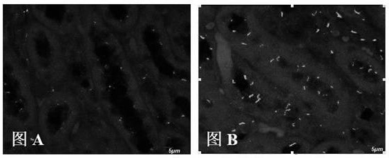
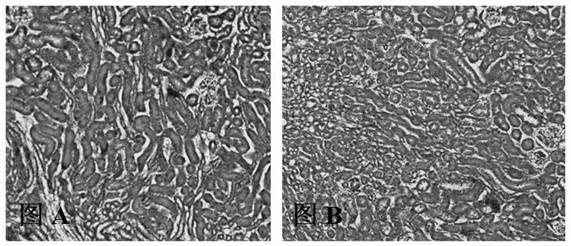
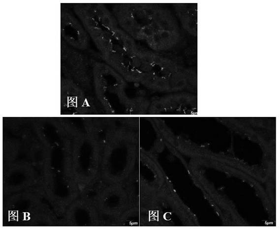
[0021] The traditional simple mouse whole body perfusion fixation, that is, after deep anesthesia, the chest cavity is opened, a small needle is inserted into the left ventricle, and at the same time, a hole is opened in the right atrium to let blood, and the blood vessels are washed with normal saline first, and then the fixative solution is injected. Therefore, the method is relatively simple, and it is a common method for HE staining of mouse tissues to observe the tissue morphology and structure. Therefore, most researchers will prefer the traditional simple mouse whole body perfusion fixation method when observing the morphological changes of the kidney or other immunohistochemical experiments. The applicant took the traditional simple mouse whole body perfusion fixation method as a comparative experiment example, and used the same PBS buffer s...


PUM

PropertyMeasurementUnit
Diameteraaaaaaaaaa

Abstract

The invention relates to the technical field of immunofluorescent staining of kidneys, and discloses a perfusion fixation method for effectively improving and observing immunofluorescence of primary cilia in the kidney of a mouse. The method comprises the following steps: after the abdominal cavity of the mouse is anesthetized, quickly opening the chest to expose the heart of the mouse, opening the left heart tip, inserting a perfusion needle connected with a peristaltic pump into the left ventricle from the opening, perfusing a certain amount of PBS for cleaning and a certain amount of 10% neutral formalin for fixing on the whole body at a constant speed at a flow speed of 40-60 [mu]l/s, taking out the kidney of the mouse when the whole body of the mouse is stiff, and fixing the kidney in a 10% neutral formalin solution for a certain time. It is found that the quantity distribution of the primary cilia of the kidney of the mouse after constant-speed perfusing is greatly improved; and compared with traditional simple whole-body tissue perfusion fixation, the cell proportion of the primary cilia is improved from 55% to 90-98%, and the correctness of a test for researching the pathogenesis of ciliary lesion diseases is greatly improved. Meanwhile, according to the above technical scheme, experimental operation repeatability is good, and a good foundation is laid for follow-up study on pathogenesis of ciliary lesion diseases.

Description

technical field [0001] The invention relates to the technical field of kidney immunofluorescence staining, in particular to a method for perfusion and fixation of mouse whole body tissue for improving the observation of mouse kidney primary cilia immunofluorescence. Background technique [0002] Cell primary cilia are dynamic microtubule structures protruding from the plasma membrane of the cell, which can act as cell sensors, adapt to changes in the extracellular environment, and maintain normal physiological functions of cells. In order to study the pathogenesis of cell native cilia disease, it needs to be fixed and stained in order to observe the cell native cilia. In the experimental study of paraffin section immunohistochemistry, the distribution and number of primary cilia in mouse kidney tissue can only be observed by immunohistochemical staining. In the experimental study of immunohistochemistry, the effect of tissue perfusion fixation is crucial to the experimental ...

Claims


Application Information

Patent Timeline
IPC IPC(8): A01N1/02
CPCA01N1/0247
Inventor 刘志强姚鑫李媛媛王振辉邢秀玲钱深
Owner HENAN POLYTECHNIC UNIV
Why Eureka
Try Eureka