Vertebra positioning and spine segmentation method based on deep learning in medical image

A medical image and deep learning technology, applied in the field of medical CT image processing, can solve problems such as poor relationship description performance, achieve the effect of improving accuracy, increasing perception ability, and improving accuracy

Active Publication Date: 2021-10-15
TONGJI UNIV
View PDF10 Cites 4 Cited by
  • Summary
  • Abstract
  • Description
  • Claims
  • Application Information

AI Technical Summary

Problems solved by technology

[0003] The purpose of the present invention is to provide a medical image based on deep learning Vertebrae Localization and Spine Segmentation Methods

Method used


Image

  • Vertebra positioning and spine segmentation method based on deep learning in medical image
  • Vertebra positioning and spine segmentation method based on deep learning in medical image
  • Vertebra positioning and spine segmentation method based on deep learning in medical image

Examples


Embodiment 1

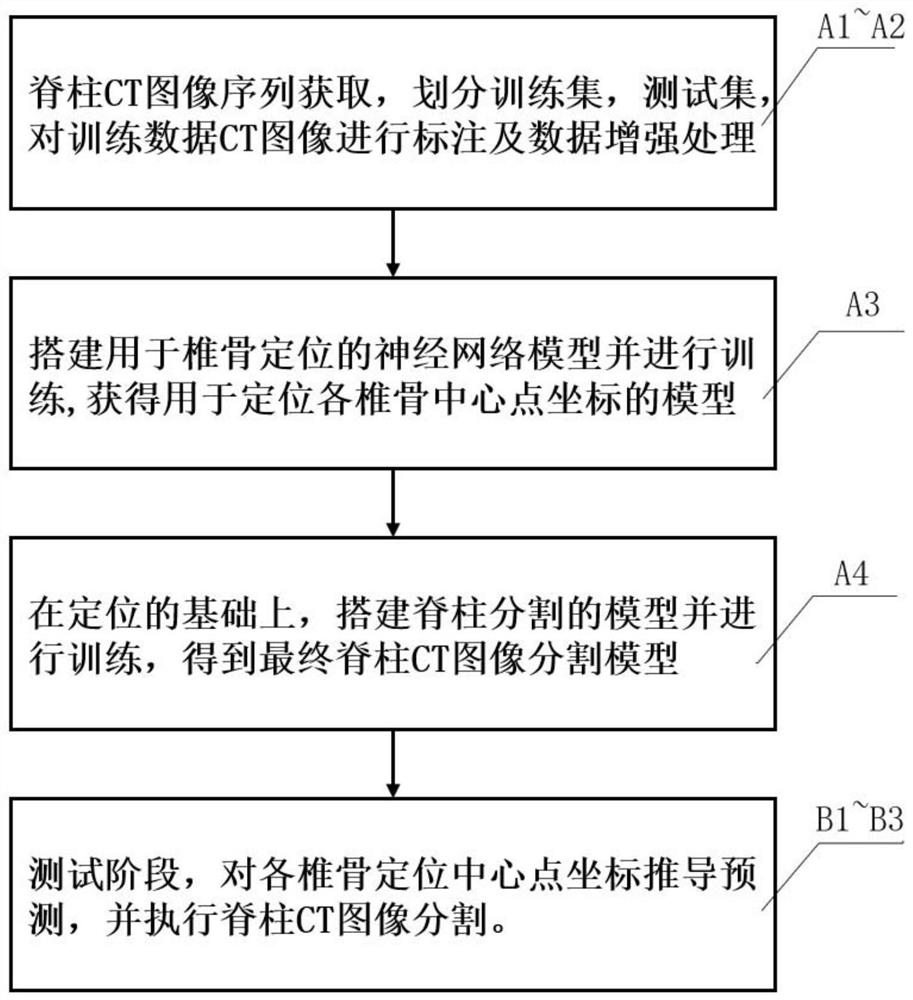
[0046] like figure 1 As shown, this embodiment provides a deep learning-based vertebral positioning and spine segmentation method in medical CT images, the method includes two stages of training and testing;

[0047] The training phase includes the following steps:

[0048] A1: training data acquisition, the training data refers to spine CT image sequences with real labels;

[0049] A2: Preprocessing the training data CT images, that is, data enhancement processing;

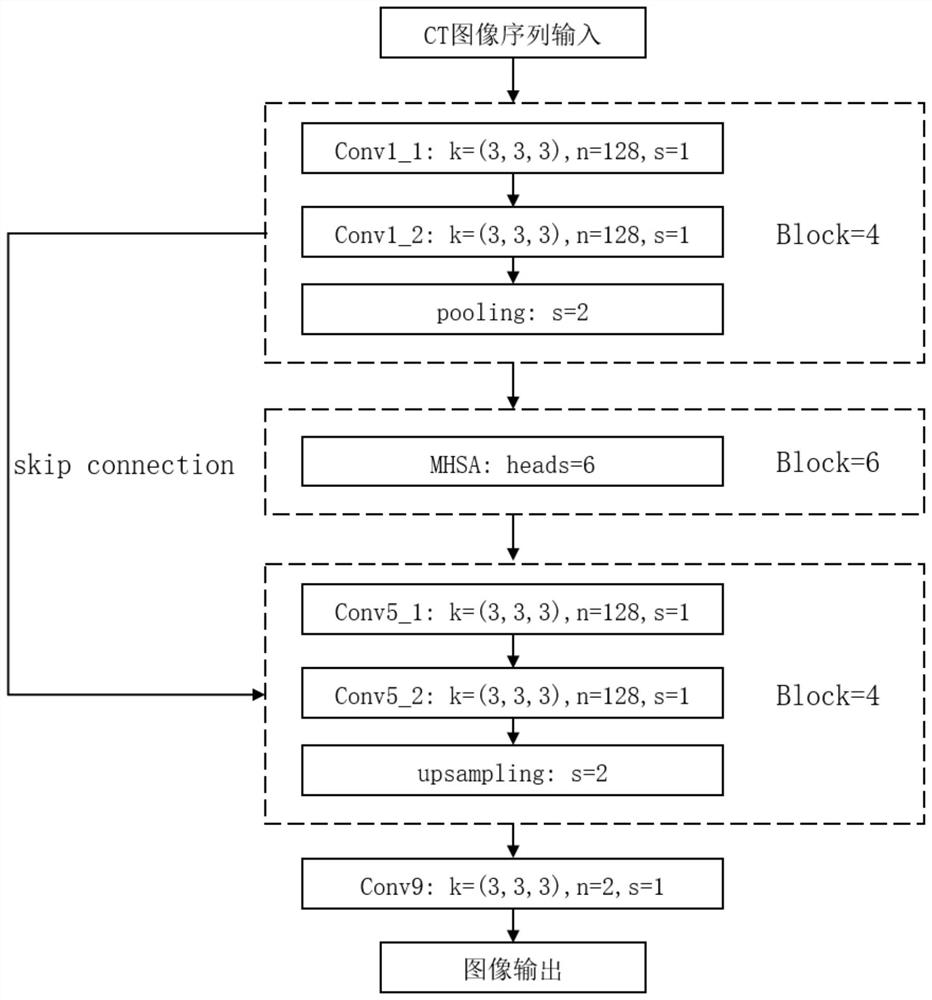
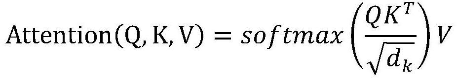
[0050] A3: Build a neural network model for vertebral positioning and perform training to obtain a model for locating the coordinates of the center points of each vertebrae;

[0051] A4: On the basis of positioning, build a spine segmentation model and perform training to obtain the final spine CT image segmentation model;

[0052] The testing phase includes the following steps:

[0053] B1: test data acquisition, the test data refers to the unlabeled spine CT image sequence to be segmented;

[0054] B2: Der...


PUM


Abstract

The invention relates to a vertebra positioning and spine segmentation method based on deep learning in a medical image, which comprises a training stage and a testing stage, and is characterized in that the training stage comprises the following steps: training data is acquired and marked; performing data enhancement processing on the training data; training a network model for vertebra positioning; on the basis of positioning, training a spine segmentation model. The test stage comprises the following steps: acquiring test data; deriving and predicting the coordinates of each vertebra positioning center point; executing spinal CT image segmentation, wherein the vertebra positioning network model and the spine segmentation model both adopt U-Net models in which a multi-head attention mechanism is introduced. Compared with the prior art, the spine CT image automatic segmentation and vertebra marking positioning are realized, the segmentation accuracy is improved by introducing a multi-head self-attention mechanism, and decision-making help is provided for subsequent complex medical diagnosis.

Description

technical field [0001] The invention relates to the field of medical CT image processing, in particular to a deep learning-based vertebral positioning and spine segmentation method in medical images. Background technique [0002] The spine is an important part of the musculoskeletal system of the human body. It plays an important role in our daily activities and load transfer while maintaining the structure of the human body and its organs. Computed tomography (CT) and magnetic resonance imaging (MRI) medical imaging technologies are now the two main means of diagnosis and treatment in spine surgery, and are widely used in screening and diagnosis for clinical and research purposes. Locating and segmenting vertebral bodies from spine CT data is a critical step in many clinical applications involving the spine, such as pathological diagnosis, surgical planning, and postoperative evaluation. The imaging mechanism of CT images is based on X-ray transmission. Considering the imp...

Claims


Application Information

Patent Timeline
Patent Type & Authority Applications(China)
IPC IPC(8): G06T7/11G06T7/70G06K9/62G06N3/04G06N3/08
CPCG06T7/11G06T7/70G06N3/08G06T2207/10081G06T2207/20081G06T2207/30012G06N3/045G06F18/214
Inventor 毛孝鑫郝泳涛
Owner TONGJI UNIV
Why Eureka
Try Eureka