
Method and system for automatically identifying microscopic image of metaphase splitting phase of chromosome

A microscopic image and automatic identification technology, which is applied in the field of radiation biological dosimetry, can solve the problems of subjectivity, time-consuming and labor-intensive manual methods, and achieve standardization and unification, and improve the recognition speed and recognition accuracy.

Active Publication Date: 2021-10-22
BEIJING HUIRONGHE TECH
View PDF5 Cites 6 Cited by
  • Summary
  • Abstract
  • Description
  • Claims
  • Application Information

AI Technical Summary

Problems solved by technology

[0003] At present, most of the related technologies use the method of manual reading under the microscope to identify dicentric chromosomes. The manual method is time-consuming and labor-intensive and is easily affected by human subjectivity.

Method used


Image

  • Method and system for automatically identifying microscopic image of metaphase splitting phase of chromosome
  • Method and system for automatically identifying microscopic image of metaphase splitting phase of chromosome
  • Method and system for automatically identifying microscopic image of metaphase splitting phase of chromosome

Examples


Embodiment Construction

[0026] In order to make the purpose, technical solution and advantages of the present invention clearer, the technical solution of the present invention will be clearly and completely described below in conjunction with the accompanying drawings of the embodiments of the present invention. Apparently, the described embodiment is one embodiment of the present invention, but not all of them. Based on the described embodiments of the present invention, all other embodiments obtained by persons of ordinary skill in the art without creative efforts shall fall within the protection scope of the present invention.

[0027] Unless otherwise defined, the technical terms or scientific terms used in the present invention shall have the usual meanings understood by those skilled in the art to which the present invention belongs.

[0028] The embodiment of the present application firstly provides an automatic identification method for microscopic images of chromosome metaphase based on con...


PUM


Abstract

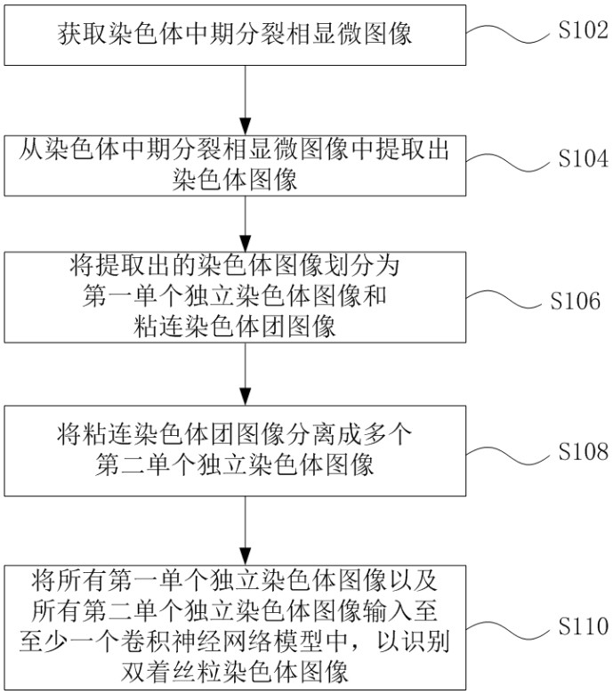
The invention discloses an automatic identification method and system for a chromosome metaphase splitting phase microscopic image based on a convolutional neural network. The automatic identification method comprises the following steps: acquiring the chromosome metaphasesplitting phase microscopic image; extracting a chromosome image from the chromosome metaphase splitting phase microscopic image; dividing the extracted chromosome image into a first single independent chromosome image and an adhesion chromosome group image; separating the adhesion chromosome group image into a plurality of second single independent chromosome images; and inputting all the first single independent chromosome images and all the second single independent chromosome images into at least one convolutional neural network model to identify a double centromere chromosome image. The double-centromere chromosome image is identified through the convolutional neural network model, so that automation in the identification process of the double-centromere chromosome is realized.

Description

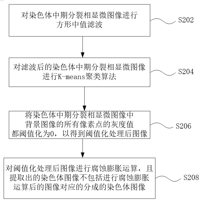
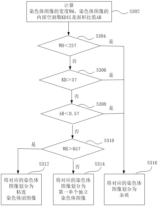
technical field [0001] The present application relates to the field of radiation biological dosimetry, in particular to a method and system for automatic recognition of microscopic images of chromosome metaphase. Background technique [0002] Chromosomal dicentric analysis (for example, of human peripheral blood lymphocytes) is the most internationally recognized and reliable method for estimating radiation biological dose. [0003] At present, most of the related technologies use manual microscopic reading to identify dicentric chromosomes, which is time-consuming and labor-intensive and is easily affected by human subjectivity. Contents of the invention [0004] In view of the above problems, the present application is proposed in order to provide a method and system for automatic identification of microscopic images of chromosome metaphase that overcome the above problems or at least partially solve the above problems. [0005] According to one aspect of the present in...

Claims


Application Information

Patent Timeline
Patent Type & Authority Applications(China)
IPC IPC(8): G06K9/00G06K9/62G06N3/04G06T7/00
CPCG06T7/0012G06T2207/10056G06T2207/20081G06T2207/20084G06T2207/30024G06N3/047G06N3/045G06F18/23213G06F18/2415
Inventor 申相周正干马腾飞李朝文杨富城温占波
Owner BEIJING HUIRONGHE TECH
Learn More