Cancellous bone modeling method, device, storage medium and electronic equipment
A cancellous bone and model technology, applied in the field of medical image processing, can solve the problems of high cost, inability to be widely used, large radiation dose, etc., and achieve the effect of high applicability
- Summary
- Abstract
- Description
- Claims
- Application Information
AI Technical Summary
Problems solved by technology
Method used
Image

Examples
Embodiment 1
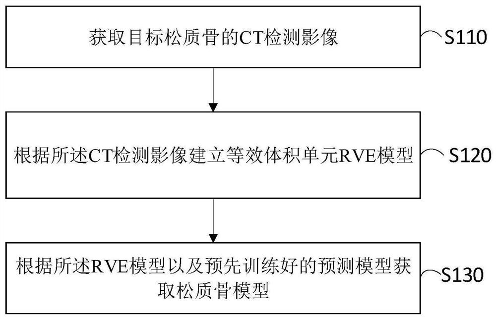
[0084] The present invention provides a cancellous bone modeling method, please refer to figure 1 , the method includes the following steps:
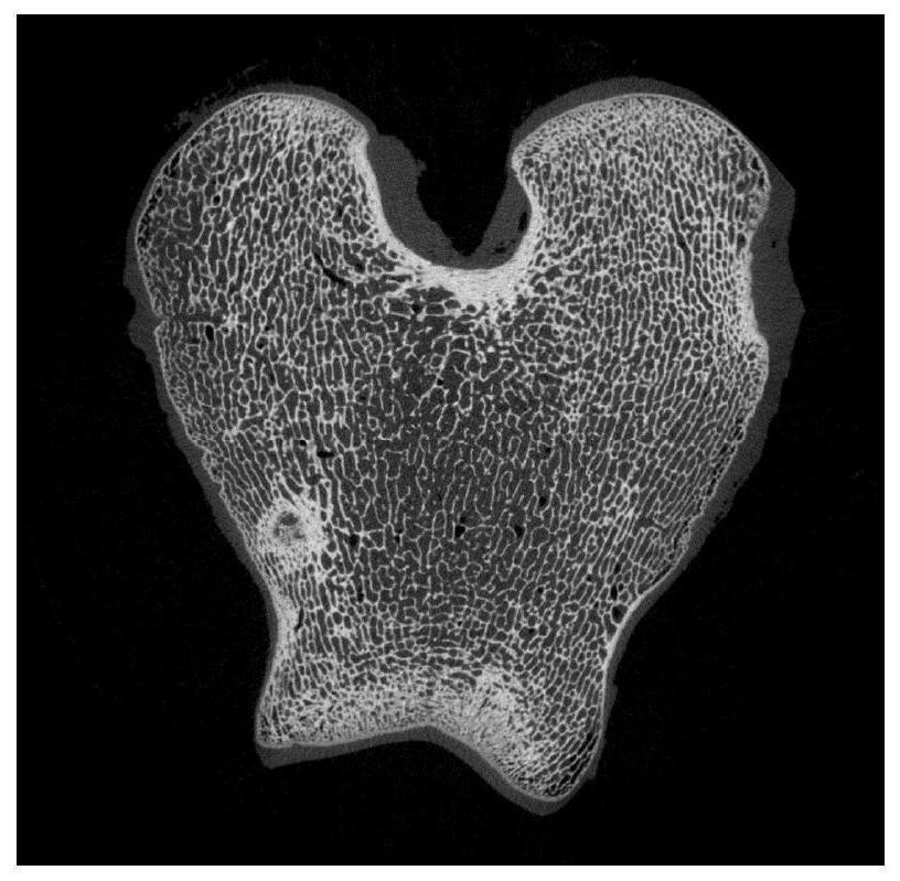
[0085] Step S110: Obtain a CT detection image of the target cancellous bone.
[0086] The target cancellous bone is the cancellous bone to be tested, and the CT detection image of the target cancellous bone is obtained by scanning a clinical TC image with a relatively low resolution. For example, the commonly used clinical CT technology for clinical observation of bone density differences has a small radiation dose, and the resolution of the CT detection image obtained by scanning is up to 0.6 mm.
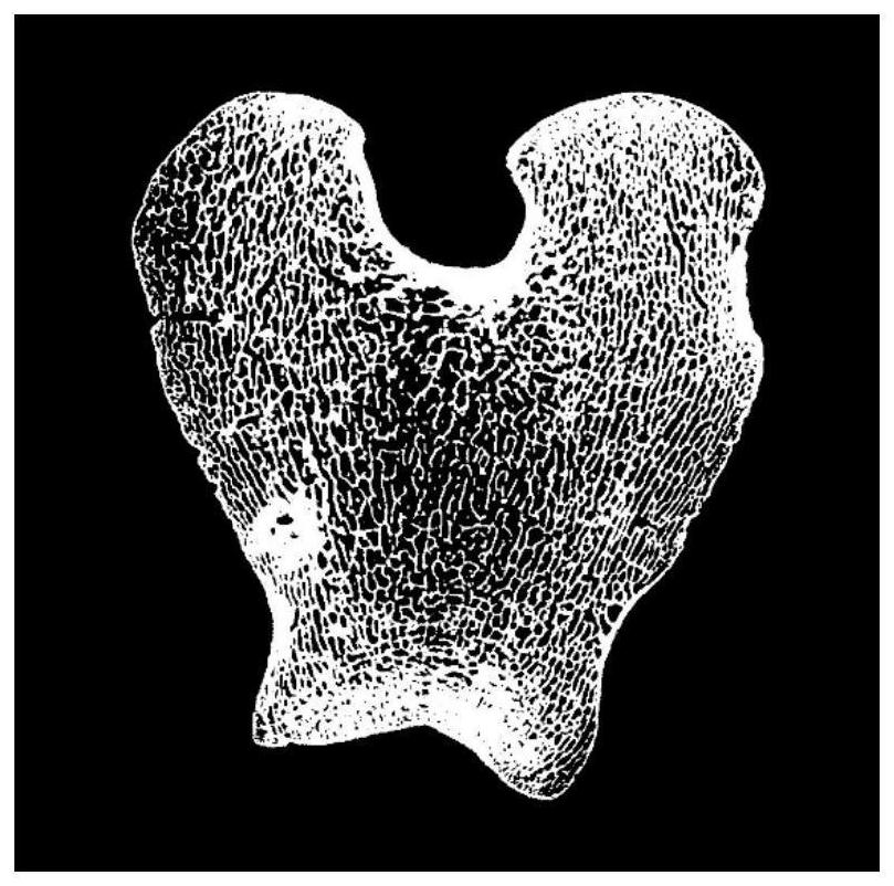
[0087] Step S120: Establish an equivalent volume unit RVE model according to the CT detection image.
[0088] The equivalent volume element RVE model is also called the representative volume element (Representative VolumeElement, RVE) model. The RVE model consists of a large number of strengthening phases and matrix phases. Its scale is h...
Embodiment 2
[0095] On the basis of the first embodiment, this embodiment describes the method in the first embodiment through specific implementation cases.
[0096] In the above cancellous bone modeling method, before obtaining the cancellous bone model according to the RVE model and the pre-trained prediction model, the method also includes the following process: firstly obtain CT images of a plurality of sample bones, and Obtain the data of the plurality of sample bones. Among them, the multiple sample bones need to include samples of different genders, different parts and different ages, and the data of multiple sample bones are obtained from the database at the same time. The data of the sample bone include the elastic modulus of the trabecular bone of the sample bone and the mapping relationship between the elastic modulus of the trabecular bone and the unit circle for conformal mapping. Then, a three-dimensional finite element model and a three-dimensional pixel information matrix...
Embodiment 3
[0099] On the basis of the second embodiment, this embodiment describes the method in the first embodiment through specific implementation cases.
[0100] Before obtaining the data of the plurality of sample bones from the database, the database needs to be obtained first, which can be obtained according to the following method. Firstly, microscopic and nano-indentation experiments are carried out on the experimental sections of the plurality of sample bones to obtain the elastic modulus of trabecular bone corresponding to each of the sample bones, and then the trabecular bone modulus of the experimental sections of each of the sample bones is obtained. The mapping relationship between the elastic modulus of the beam and the unit circle is conformally mapped, and finally a database including the elastic modulus of the trabecular bone and the mapping relationship can be obtained.
[0101] Specifically, firstly, according to the conformal mapping method, the mapping relationship...
PUM
 Abstract
Description
Claims
Application Information
 - R&D
- Intellectual Property
- Life Sciences
- Materials
- Tech Scout
- Unparalleled Data Quality
- Higher Quality Content
- 60% Fewer Hallucinations
Browse by: Latest US Patents, China's latest patents, Technical Efficacy Thesaurus, Application Domain, Technology Topic, Popular Technical Reports.
© 2025 PatSnap. All rights reserved.Legal|Privacy policy|Modern Slavery Act Transparency Statement|Sitemap|About US| Contact US: help@patsnap.com