
Ultrasonic imaging method and system

An ultrasound imaging and ultrasound image technology, applied in the field of medical devices, can solve the problems of large reading volume, misdiagnosis, missed diagnosis, omission or forgetting of important information, etc., to achieve the effect of improving work efficiency and improving accuracy

Pending Publication Date: 2021-11-26
SHENZHEN MINDRAY BIO MEDICAL ELECTRONICS CO LTD
View PDF0 Cites 0 Cited by
  • Summary
  • Abstract
  • Description
  • Claims
  • Application Information

AI Technical Summary

Problems solved by technology

If the patient’s condition is more complicated and the amount of reading is relatively large, when there are many patients and clinicians are under high work pressure, especially in the case of multiple lesions, important information may be missed or forgotten, which may easily lead to misdiagnosis and missed diagnosis

Method used


Image

  • Ultrasonic imaging method and system
  • Ultrasonic imaging method and system
  • Ultrasonic imaging method and system

Examples


Embodiment Construction

[0055] The present invention will be further described in detail below through specific embodiments in conjunction with the accompanying drawings. Wherein, similar elements in different implementations adopt associated similar element numbers. In the following implementation manners, many details are described for better understanding of the present application. However, those skilled in the art can readily recognize that some of the features can be omitted in different situations, or can be replaced by other elements, materials, and methods. In some cases, some operations related to the application are not shown or described in the description, this is to avoid the core part of the application being overwhelmed by too many descriptions, and for those skilled in the art, it is necessary to describe these operations in detail Relevant operations are not necessary, and they can fully understand the relevant operations according to the description in the specification and genera...


PUM


Abstract

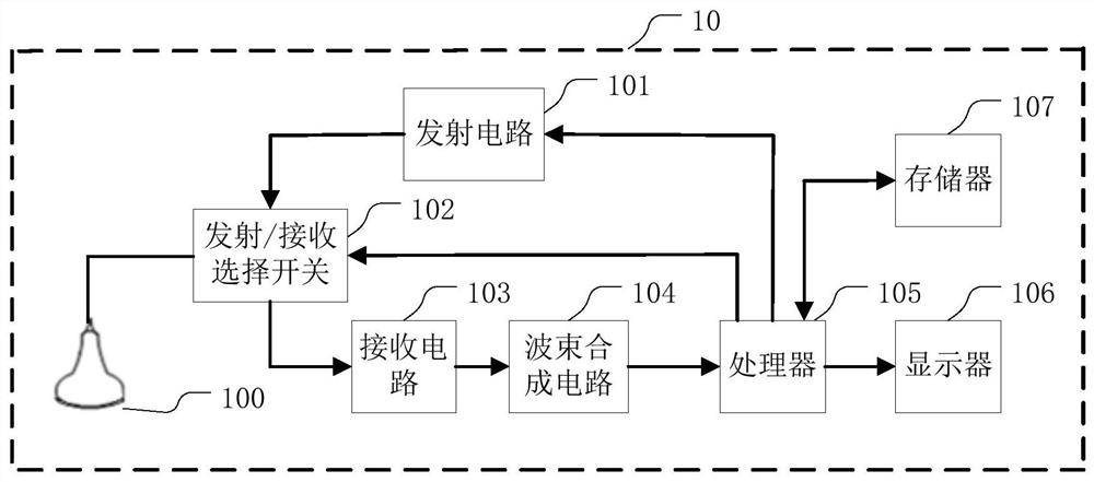
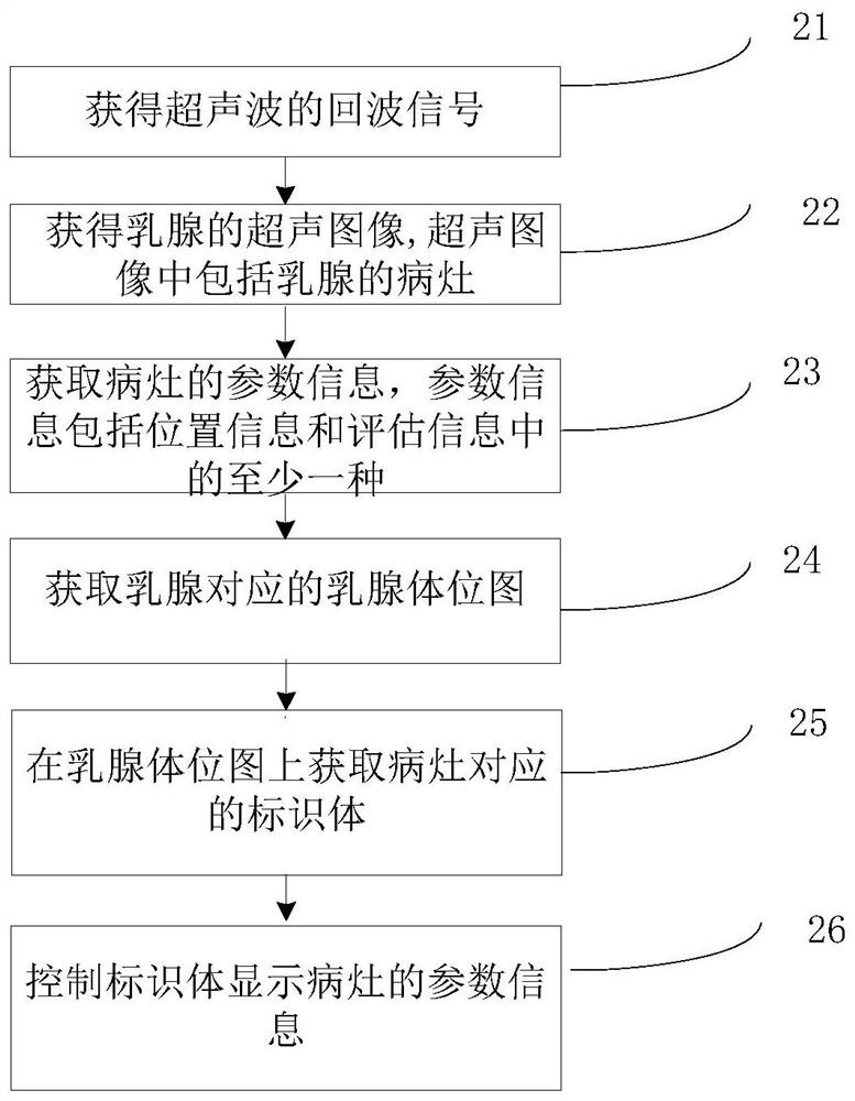
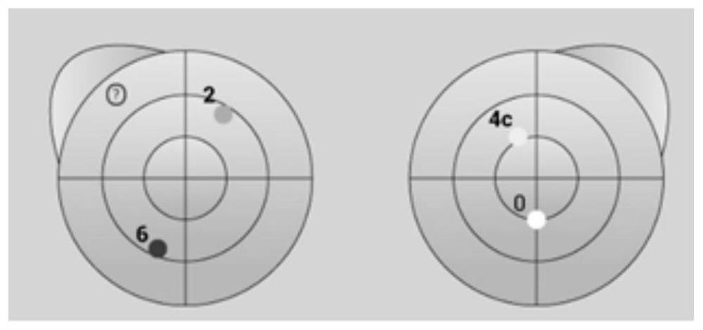
The invention relates to an ultrasonic imaging method and system. A transmitting circuit and a receiving circuit excite an ultrasonic probe to transmit ultrasonic waves to the mammary gland, receive echoes of the ultrasonic waves and obtain echo signals of the ultrasonic waves; a processor generates an ultrasonic image of the mammary gland according to the echo signals of the ultrasonic waves; the processor obtains parameter information of a lesion, wherein the parameter information comprises at least one of position information of the lesion and evaluation information of the lesion; the processor obtains a mammary gland body position map corresponding to the mammary gland; an identification body corresponding to the lesion is obtained on the mammary gland body position map; and the processor controls the identification body to display parameter information of the lesion. By displaying the parameter information of the lesion in the mammary gland body position image, an operator can obtain the information of the lesion very accurately and visually, the accuracy of obtaining the information of the lesion by the operator is improved, and the working efficiency of the operator is also improved.

Description

technical field [0001] The present application relates to the field of medical devices, in particular to an ultrasonic imaging method and system. Background technique [0002] At present, there is a lack of an intuitive display function for browsing the evaluation of breast lesions in patients as a whole in clinical practice. After receiving the ultrasound report, clinicians can only understand the results of ultrasound examination by reading text. If the patient's condition is more complicated and the amount of reading is relatively large, when there are many patients and clinicians are under high work pressure, especially in the case of multiple lesions, important information may be missed or forgotten, which may easily lead to misdiagnosis and missed diagnosis. Contents of the invention [0003] In one embodiment, a method of ultrasound imaging is provided, comprising: [0004] Exciting the ultrasound probe to transmit ultrasound to the breast, and receiving the echo o...

Claims


Application Information

Patent Timeline
Patent Type & Authority Applications(China)
IPC IPC(8): A61B8/08A61B8/00
Inventor 温博
Owner SHENZHEN MINDRAY BIO MEDICAL ELECTRONICS CO LTD
Learn More