Supercharge Your Innovation With Domain-Expert AI Agents!

Breast nodule calcification analysis device

A breast nodule and analysis device technology, applied in image analysis, mammography, organ motion/change detection, etc., to achieve the effect of convenient and accurate judgment

Pending Publication Date: 2021-12-17
什维新智医疗科技(上海)有限公司
View PDF9 Cites 0 Cited by
  • Summary
  • Abstract
  • Description
  • Claims
  • Application Information

AI Technical Summary

Problems solved by technology

Unfortunately, there have been no studies to quantify BI-RADS features to improve the diagnostic performance of breast cancer classification

Method used


Image

  • Breast nodule calcification analysis device
  • Breast nodule calcification analysis device
  • Breast nodule calcification analysis device

Examples


Embodiment Construction

[0039] Below in conjunction with specific embodiment, further illustrate the present invention. It should be understood that these examples are only used to illustrate the present invention and are not intended to limit the scope of the present invention. In addition, it should be understood that after reading the teachings of the present invention, those skilled in the art can make various changes or modifications to the present invention, and these equivalent forms also fall within the scope defined by the appended claims of the present application.

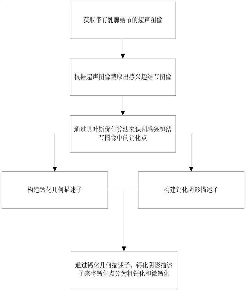
[0040] Embodiments of the present invention relate to a breast nodule calcification analysis device, please refer to figure 1 ,include:

[0041] Image acquisition module: used to acquire ultrasound images with breast nodules;

[0042] The nodule image extraction module of interest: for intercepting the breast nodule boundary of the ultrasound image by selecting several coordinate points of interest to obtain the nodule image ...


PUM


Abstract

The invention relates to a mammary nodule calcification analysis device. The mammary nodule calcification analysis device comprises an image acquisition module which is used for acquiring an ultrasonic image with a mammary nodule; a nodule-of-interest image extraction module used for intercepting the breast nodule boundary of the ultrasonic image by selecting a plurality of coordinate points of interest to obtain a nodule-of-interest image; a calcification recognition module used for recognizing calcification points in the nodule image of interest through a Bayesian optimization algorithm to obtain an image Icals_grow; a calcification shadow descriptor construction module used for constructing a calcification shadow descriptor according to a calcification point in the image Icals_grow; a calcification geometric descriptor construction module used for constructing a calcification geometric descriptor according to a calcification point in the image Icals_grow; and a coarse calcification and micro calcification extraction module used for classifying calcification points in the image Icals_grow into coarse calcification or micro calcification by the calcification shadow descriptor and the calcification geometric descriptor. The calcification point can be effectively analyzed.

Description

technical field [0001] The invention relates to the technical field of auxiliary medical diagnosis, in particular to a breast nodule calcification analysis device. Background technique [0002] Today, with the increasing demand for rapid and accurate diagnosis and the shortage of clinical personnel, computer analysis methods have been increasingly used to support routine clinical diagnosis and have shown good results. [0003] Breast cancer is expected to become the second most deadly cancer in women in recent years, with a mortality rate of 15%. These statistics show that a breast cancer diagnosis is critical to improving life expectancy, especially for women. As a commonly used clinical tool, ultrasound imaging is a non-invasive, radiation-free, and low-cost technique for cancer diagnosis. However, identifying breast lesions and detecting signs of cancer from ultrasound is a challenging task due to low image quality. [0004] The growth and progression of a malignant tu...

Claims


Application Information

Patent Timeline
Patent Type & Authority Applications(China)
IPC IPC(8): G06T7/00G06T7/12G06T7/187G06T7/62G06K9/32A61B8/08A61B8/00
CPCG06T7/0012G06T7/62G06T7/12G06T7/187A61B8/0825A61B8/5207A61B8/5215A61B8/5223G06T2207/20132G06T2207/30068G06T2207/30096G06T2207/10132G06T2207/20084
Inventor 韩东旭
Owner 什维新智医疗科技(上海)有限公司
Features
  • R&D
  • Intellectual Property
  • Life Sciences
  • Materials
  • Tech Scout
  • Unparalleled Data Quality
  • Higher Quality Content
  • 60% Fewer Hallucinations
Learn More