Hip joint standard ultrasonic image acquisition method based on Graf ultrasonic technology and intelligent system

A technology of ultrasonic image and ultrasonic technology, applied in the directions of ultrasonic/sonic/infrasonic image/data processing, ultrasonic/sonic/infrasonic Permian technology, ultrasonic/sonic/infrasonic diagnosis, etc. Equipment, heavy workload and other problems, to achieve the effect of improving screening accuracy, getting rid of dependence, and avoiding errors in screening

Inactive Publication Date: 2022-01-21
扬州市妇幼保健院
View PDF1 Cites 1 Cited by
  • Summary
  • Abstract
  • Description
  • Claims
  • Application Information

AI Technical Summary

Problems solved by technology

In addition, as an academic research, there are differences between the Graf method and other DDH diagnostic methods. The main point of controversy is that the Graf method does not have a corresponding relationship with the physical structure of the hip joint, so the measurement results cannot be used to explain the three-dimensional structure of the hip joint.
[0011] (2) The equipment standard of the Graf method is not perfect, and the screening technical means and methods are not easy to master
The lack of effective equipment has brought great troubles to doctors
[0012] (3) Carrying out a comprehensive Graf method for hip joint ultrasound screening is a heavy workload, and it is necessary to solve the ...

Method used


Image

  • Hip joint standard ultrasonic image acquisition method based on Graf ultrasonic technology and intelligent system
  • Hip joint standard ultrasonic image acquisition method based on Graf ultrasonic technology and intelligent system
  • Hip joint standard ultrasonic image acquisition method based on Graf ultrasonic technology and intelligent system

Examples


Embodiment Construction

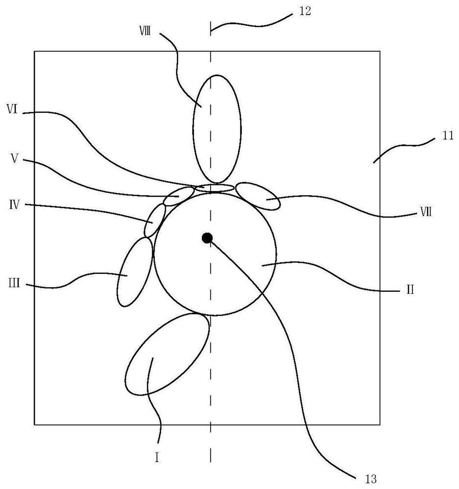
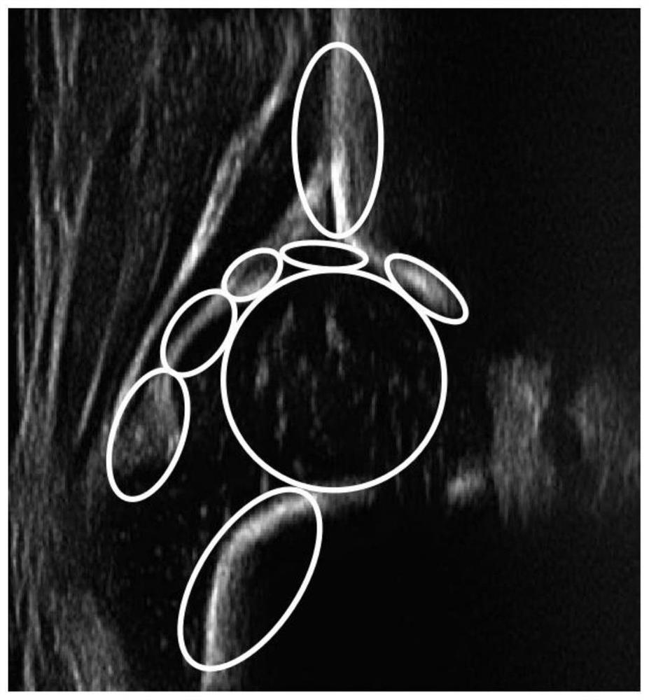
[0079] Before describing the technical scheme of the present invention in detail, the attached Figure 5-7 First, the positional relationship in the process of acquiring related ultrasound images is described as follows: the baby is lying on the examination bed, and the ultrasound probe 2 is set on the upper part of the baby’s upward hip joint (generally, it needs to be gently fitted, given that the existing technology is not suitable for driving ultrasound). Gentle fitting of the probe type 2 actuator and the human body is a mature technology, which will not be described in detail in the present invention). The axial direction of the ultrasonic probe 2 is set as the X direction, the width direction of the ultrasonic probe 2 (that is, the head and tail direction of the crib) is set as the Z direction; direction) is the Y direction; in Figure 5 The right part of the middle ultrasonic probe 2 shows the position of the base 11 of the picture, where the online ultrasonic image i...


PUM


Abstract

The invention discloses a hip joint standard ultrasonic image acquisition method based on a Graf ultrasonic technology and an intelligent system, and relates to an intelligent medical equipment system, in particular to an early screening technology for infant developmental dysplasia of joints. The method comprises the following steps: 1) establishing a picture substrate with a baseline; 2) presetting an identification area and an identification condition on the picture substrate; 3) acquiring an online ultrasonic image, and comparing and judging the online ultrasonic image with the preset identification area and the preset identification condition on the picture substrate; and 4) generating a standard image which can be used for measurement by using the online ultrasonic image which falls into the preset identification area and meets the preset identification condition. In the step 1), the ratio of the width to the height of the picture substrate is 1: (1-1.3), and a base line is arranged in the range of 10-20% of the width size of the two sides of the central point in the longitudinal direction of the picture substrate. According to the method, errors in screening can be effectively avoided, and the screening accuracy and efficiency are greatly improved.

Description

technical field [0001] The invention relates to an intelligent medical equipment system, in particular to an early screening technology for infant developmental dysplasia of the hip. Background technique [0002] Infant developmental dysplasia of hip (DDH) is birth-related and refers to abnormalities in the structure of the hip joint at birth and progressive deterioration during development, including hip joint instability to hip dislocation series of lesions. DDH ranks fifth among birth defects and is one of the most common musculoskeletal disorders that seriously endanger the health of infants. The etiology of DDH is unknown, but it may be related to genetics, amniotic fluid, fetal position, and birth conditions. Epidemiological studies have shown that about 12% to 33% of children with DDH have a positive family history, and the incidence of mechanical factors such as breech delivery is 16%; the incidence rate of girls is much higher than that of boys, and the risk facto...

Claims


Application Information

Patent Timeline
IPC IPC(8): A61B8/00A61B8/08
CPCA61B8/4209A61B8/5215A61B8/0875
Inventor 王加宽
Owner 扬州市妇幼保健院
Why Eureka
Try Eureka