
Method for detecting abnormal blood vessel structure or form in blood vessel image and related product

A technology of abnormal shape and vascular structure, applied in the field of medical imaging, can solve the problems of low detection efficiency and achieve the effect of improving efficiency and detection efficiency

Pending Publication Date: 2022-01-28
SHANGHAI UNITED IMAGING INTELLIGENT MEDICAL TECH CO LTD
View PDF0 Cites 2 Cited by
  • Summary
  • Abstract
  • Description
  • Claims
  • Application Information

AI Technical Summary

Problems solved by technology

Therefore, the traditional variation detection method of vascular rings has the problem of low detection efficiency.

Method used


Image

  • Method for detecting abnormal blood vessel structure or form in blood vessel image and related product
  • Method for detecting abnormal blood vessel structure or form in blood vessel image and related product
  • Method for detecting abnormal blood vessel structure or form in blood vessel image and related product

Examples


Embodiment Construction

[0047] In order to make the purpose, technical solution and advantages of the present application clearer, the present application will be further described in detail below in conjunction with the accompanying drawings and embodiments. It should be understood that the specific embodiments described here are only used to explain the present application, and are not intended to limit the present application.

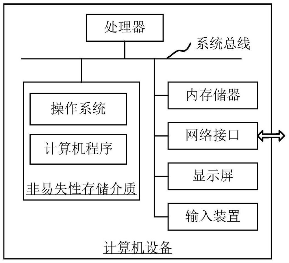
[0048] The method for detecting abnormal structure or shape of blood vessels in blood vessel images provided by the embodiment of the present application can be applied to such as figure 1 computer equipment shown. The computer device includes a processor and a memory connected through a system bus, and a computer program is stored in the memory. When the processor executes the computer program, the steps of the following method embodiments can be performed. Optionally, the computer device may also include a network interface, a display screen and an input device. Wherei...


PUM


Abstract

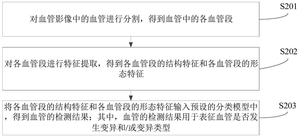
The invention relates to a method for detecting abnormal blood vessel structure or form in a blood vessel image and a related product. The method comprises the following steps: segmenting a blood vessel in a blood vessel image to obtain each blood vessel segment in the blood vessel; performing feature extraction on each blood vessel segment to obtain structural features of each blood vessel segment and morphological features of each blood vessel segment; inputting the structural features of the blood vessel segments and the morphological features of the blood vessel segments into a preset classification model to obtain a blood vessel detection result; wherein the detection result of the blood vessel is used for representing whether the blood vessel is varied or not and / or the variation type. The efficiency of detecting whether the blood vessel is mutated or not can be improved.

Description

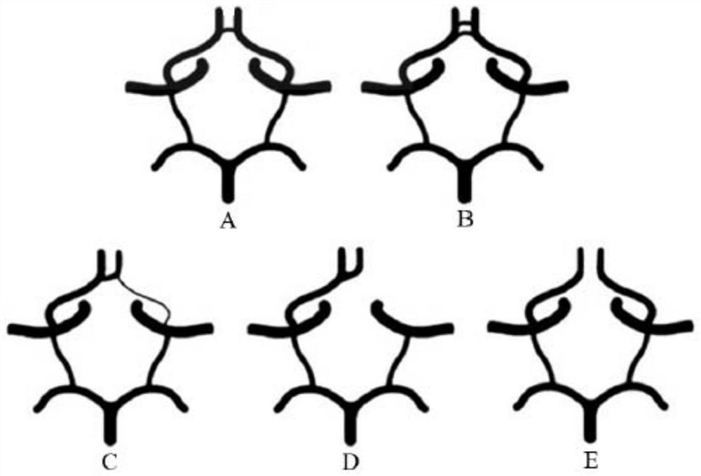
technical field [0001] The present application relates to the technical field of medical imaging, in particular to a method for detecting abnormal structure or shape of blood vessels in vascular images and related products. Background technique [0002] The vascular ring is a major collateral circulation in the blood supply system of the human body. For example, the Willis circle (Willis circle) consists of the A1 segment of the anterior cerebral artery on both sides, the anterior communicating artery, the ends of the internal carotid artery on both sides, and the P1 segment of the posterior cerebral artery on both sides. As well as the posterior communicating arteries on both sides, it is the main collateral circulation in the blood supply system of the brain, providing a communication path between the anterior circulation (carotid artery) and the posterior circulation (vertebrobasilar artery). However, the variation rate of vascular rings is high, which is manifested by th...

Claims


Application Information

Patent Timeline
IPC IPC(8): G06T7/00G06T7/11G06V10/44G06V10/764G06V10/82G06K9/62G06N3/04G06N3/08G06N20/10
CPCG06T7/0012G06T7/11G06N3/04G06N3/08G06N20/10G06T2207/10081G06T2207/10088G06T2207/20081G06T2207/20084G06T2207/30101G06F18/241G06F18/2411
Inventor 刘姗姗曹晓欢江鹏博薛忠
Owner SHANGHAI UNITED IMAGING INTELLIGENT MEDICAL TECH CO LTD
Learn More