Brain tumor detection method based on attention mechanism and MRI (Magnetic Resonance Imaging) multi-modal fusion

A detection method and attention technology, applied in the intersection of computer and medicine, in the field of deep learning, which can solve the problems of huge data volume, unclear outline, and large 3D convolution kernel.

Pending Publication Date: 2022-03-01
BEIJING UNIV OF TECH
View PDF0 Cites 4 Cited by
  • Summary
  • Abstract
  • Description
  • Claims
  • Application Information

AI Technical Summary

Problems solved by technology

The 3D convolution kernel itself has a relatively large amount of parameters, and this is only the number of one channel. If it is used to extract the source 3D MRI image and store the features in multiple channels, the overall data volume will be very large. Memory and video memory will not be able to support
The existing segmentation network, regardless of whether the model is based on 2D or 3D, can segment the general outline of the lesion area. In addition to the need to consider the connection between 2D images, there are still some areas for improvement in terms of convolution, such as in T1 , In the T1c image, most of the contours are not very clear, especially the edema area, and this phenomenon also leads to the problem of excessive segmentation when the network extracts features.

Method used


Image

  • Brain tumor detection method based on attention mechanism and MRI (Magnetic Resonance Imaging) multi-modal fusion
  • Brain tumor detection method based on attention mechanism and MRI (Magnetic Resonance Imaging) multi-modal fusion
  • Brain tumor detection method based on attention mechanism and MRI (Magnetic Resonance Imaging) multi-modal fusion

Examples


Embodiment Construction

[0056] The present invention will be described in detail below in conjunction with the accompanying drawings and embodiments.

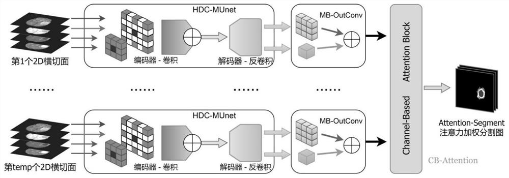
[0057] A brain tumor detection method based on attention mechanism and MRI multimodal fusion, the method comprises the following steps,

[0058] Step S1: Dataset introduction.

[0059] In order to verify the performance of the model, Brats2015 is selected as the data set. The entire data set contains 274 samples, of which 220 samples are HGG cases, medically known as high-grade glioma, which is a poorly differentiated malignant tumor; the remaining 54 samples are LGG cases, medically known as low-grade glioma Tumors, these are benign tumors with good differentiation properties. Each sample in the dataset contains 5 3D Volumes, and each 3D Volume consists of 155 layers of 2D images. The first four 3D images are MRI scan results [T1, T2, T1c, Flair], which represent the basic structure, tissue water content, tissue blood supply, and tissue bound wate...


PUM


Abstract

The invention discloses a brain tumor detection method based on attention mechanism and MRI (Magnetic Resonance Imaging) multi-modal fusion, and the method comprises the steps: replacing a common convolution block of an encoder with a mixed cavity convolution block based on a Multi-Unet model; a multi-branch output convolution block MB-OutConv for short is autonomously designed by referring to a multi-branch encoder structure of an Inception model; designing an attention module CB-Attention based on channels, capturing pixel point association among the channels of the original segmented image, and performing attention weighting on the channels; a neural network is properly improved, a new attention module is independently designed to further perfect a segmentation result, and the attention module is based on an image channel and completes attention weighting at a pixel point level. And finally, segmenting the tumor and other lesion areas in the brain MRI image. On the basis of a multi-modal convolutional neural network Multi-Unet, part of encoder branches are improved, and an attention module is added behind the multi-modal convolutional neural network Multi-Unet to jointly improve the segmentation effect of the brain tumor.

Description

technical field [0001] The invention relates to deep learning, the field of medical image processing, and is an interdisciplinary field of computers and medicine. Based on the multi-modal convolutional neural network, combined with the current challenges, the neural network is appropriately improved, and a new attention module is independently designed to further improve its segmentation results. Attention weighting is done at the point level. The ultimate goal is to segment tumors and other diseased regions in brain MRI images. Background technique [0002] Image segmentation is the process of covering the target contour with a pixel-level mask. Since the shape of the lesion area is irregular, the doctor must judge the location of the disease as accurately as possible when diagnosing, otherwise, the healthy tissue will be damaged. Surgery is performed as a diseased area, which will put the patient's life in danger, so image segmentation is more meaningful than target dete...

Claims


Application Information

Patent Timeline
Patent Type & Authority Applications(China)
IPC IPC(8): G06T7/00G06V10/26G06V10/764G06K9/62G06N3/04
CPCG06T7/0012G06T2207/10088G06T2207/30016G06T2207/30096G06N3/045G06F18/2431
Inventor 蒋宗礼李聪张津丽顾问
Owner BEIJING UNIV OF TECH
Why Eureka
Try Eureka