Dual-mode coronary vessel image three-dimensional fusion method and fusion system

A vascular image, three-dimensional fusion technology, applied in the direction of ultrasound/sonic/infrasonic image/data processing, catheter, ultrasound/sonic/infrasonic Permian technology, etc., can solve inaccurate results, do not consider catheter bending and twisting, etc. problem, to achieve the effect of high resolution and deep detection depth

Active Publication Date: 2022-03-08
天津恒宇医疗科技有限公司
View PDF9 Cites 6 Cited by
  • Summary
  • Abstract
  • Description
  • Claims
  • Application Information

AI Technical Summary

Problems solved by technology

Since IVUS and OCT itself cannot provide spatial geometric information for each image frame, this method does not tak

Method used


Image

  • Dual-mode coronary vessel image three-dimensional fusion method and fusion system
  • Dual-mode coronary vessel image three-dimensional fusion method and fusion system
  • Dual-mode coronary vessel image three-dimensional fusion method and fusion system

Examples


Embodiment Construction

[0051] The present invention will be further described in detail through the accompanying drawings and specific embodiments below.

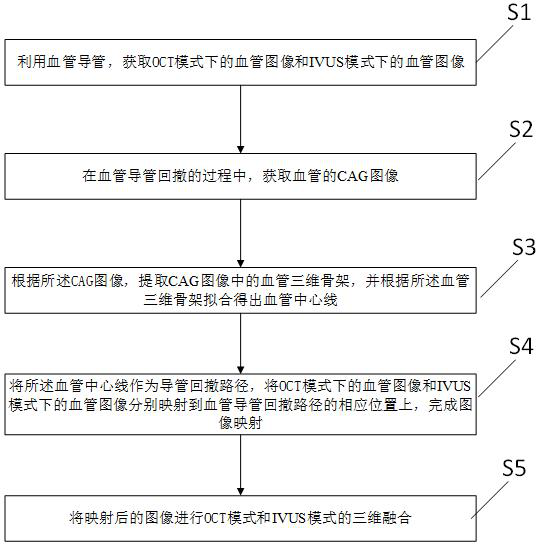
[0052] Such as figure 1 As shown, a method for three-dimensional fusion of a dual-mode coronary artery image provided by an embodiment of the present invention includes the following steps:

[0053] S1. Using a vascular catheter, obtain a vascular image in OCT mode (hereinafter referred to as OCT image) and a vascular image in IVUS mode (hereinafter referred to as IVUS image);

[0054] S2. During the process of withdrawing the vascular catheter, acquire a CAG image of the blood vessel;

[0055] Further preferably, in S1, when acquiring the vascular images of the vascular catheter in the OCT mode and the vascular images in the IVUS mode, it includes a synchronous acquisition mode and an asynchronous switching mode; the synchronous acquisition mode is the simultaneously acquired OCT mode The working mode of the blood vessel image in the IVUS mode...


PUM


Abstract

The invention discloses a dual-mode coronary blood vessel image three-dimensional fusion method and fusion system, and the method comprises the steps: obtaining a blood vessel image in an OCT mode, a blood vessel image in an IVUS mode, and a CAG image of a blood vessel through a blood vessel catheter; extracting a blood vessel three-dimensional skeleton in the CAG image according to the CAG image, and fitting according to the blood vessel three-dimensional skeleton to obtain a blood vessel center line; taking the center line of the blood vessel as a catheter withdrawing path, and respectively mapping the blood vessel image in the OCT mode and the blood vessel image in the IVUS mode to corresponding positions of the catheter withdrawing path of the blood vessel to complete image mapping; performing three-dimensional fusion of an OCT mode and an IVUS mode on the mapped image; the advantages of the three-dimensional CAG image in space, the strong tissue penetrating power of the IVUS and the high resolution of the OCT are fully exerted, more comprehensive information of the blood vessel wall and the coronary atherosclerotic plaque can be obtained, and therefore a more effective basis is provided for computer-assisted diagnosis and treatment of the coronary heart disease, evaluation of the interventional therapy effect and the like.

Description

technical field [0001] The invention relates to the technical field of blood vessel imaging, in particular to a method for three-dimensional fusion of dual-mode coronary blood vessel images. Background technique [0002] Globally, cardiovascular and cerebrovascular diseases have become one of the major diseases that threaten human health, and coronary atherosclerosis is the main cause of cardiovascular and cerebrovascular diseases. X-ray coronary angiography (CAG), intravascular ultrasound (IVUS) and optical coherence tomography (OCT) are currently the main imaging techniques for the diagnosis of cardiovascular and cerebrovascular diseases. This imaging technology can only provide two-dimensional plane image information, and cannot intuitively reflect the actual spatial situation. Due to the limitations of 2D images, 3D reconstruction and visualization of medical images has become a research hotspot. [0003] CAG judges the location, nature and degree of abnormal vascular ...

Claims


Application Information

Patent Timeline
IPC IPC(8): A61B5/00A61B8/08A61B8/12
CPCA61B5/0066A61B8/0891A61B8/12A61B8/5246
Inventor 武西宁赵士勇
Owner 天津恒宇医疗科技有限公司
Why Eureka
Try Eureka