Brain image-based infarction region segmentation method and device, equipment and medium

A region segmentation and region technology, applied in the field of image processing, can solve the problem of inability to accurately identify the actual volume of cerebral infarction, and achieve the effect of enhancing versatility and efficiency

Active Publication Date: 2022-04-01
沈阳东软智能医疗科技研究院有限公司
View PDF3 Cites 1 Cited by
  • Summary
  • Abstract
  • Description
  • Claims
  • Application Information

AI Technical Summary

Problems solved by technology

[0004] However, since brain images belong to CT single-mode images, and the cerebrospinal fluid in the ventricle has the same performance as the infarct lesion on certain two perfusion parameters represented in the brain images, the gray scale of the cerebrospinal fluid and infarct area in the brain images The actual volume of the brain infarction in the brain image cannot be accurately identified due to the presence of adhesions in three-dimensional space

Method used


Image

  • Brain image-based infarction region segmentation method and device, equipment and medium
  • Brain image-based infarction region segmentation method and device, equipment and medium
  • Brain image-based infarction region segmentation method and device, equipment and medium

Examples


Embodiment Construction

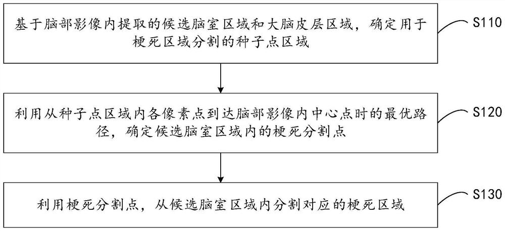
[0030] The following will clearly and completely describe the technical solutions in the embodiments of the present invention with reference to the accompanying drawings in the embodiments of the present invention. Obviously, the described embodiments are only some, not all, embodiments of the present invention. Based on the embodiments of the present invention, all other embodiments obtained by persons of ordinary skill in the art without making creative efforts belong to the protection scope of the present invention.

[0031] It should be noted that the terms "first" and "second" in the description and claims of the present invention and the above drawings are used to distinguish similar objects, but not necessarily used to describe a specific sequence or sequence. It is to be understood that the data so used are interchangeable under appropriate circumstances such that the embodiments of the invention described herein can be practiced in sequences other than those illustrate...


PUM


Abstract

The invention provides an infarction region segmentation method and device based on a brain image, equipment and a medium. The method comprises the steps of determining a seed point region for infarction region segmentation based on a candidate ventricle region and a cerebral cortex region extracted from a brain image; determining an infarction segmentation point in the candidate ventricle region by using an optimal path from each pixel point in the seed point region to a central point in the brain image; and segmenting a corresponding infarction region from the candidate ventricle region by using the infarction segmentation point. According to the method, the ant colony algorithm is adopted to analyze the infarction segmentation points in the candidate ventricle region, so that the corresponding infarction region is segmented from the candidate ventricle region by using the infarction segmentation points, the infarction region adhered to cerebrospinal fluid in the candidate ventricle region is automatically and effectively segmented, and the universality and efficiency of infarction region segmentation are enhanced.

Description

technical field [0001] The embodiments of the present application relate to the technical field of image processing, and in particular to a brain image-based infarction region segmentation method, device, device, and medium. Background technique [0002] Acute ischemic cerebrovascular disease, also known as acute ischemic stroke, is a major disease that seriously threatens the health of our population and hinders social and economic development, with high disability and fatality rates. [0003] In the treatment of acute ischemic cerebrovascular disease, computerized tomography perfusion imaging (CTP) can usually be used to conduct continuous CT scans of the patient's brain at different levels, so as to obtain the corresponding brain video. Then, according to the atlas of various perfusion parameters represented in the scanned brain image, it is judged whether there is a cerebral infarction, and then the corresponding cerebral infarction area is determined. [0004] However...

Claims


Application Information

Patent Timeline
Patent Type & Authority Applications(China)
IPC IPC(8): G06T7/11G06T7/187
Inventor 马丽娟张霞蔡巍
Owner 沈阳东软智能医疗科技研究院有限公司
Why Eureka
Try Eureka