
Medical image processing method and device

A medical image and processing method technology, applied in the computer field, can solve the problems of low image loss quality, lack of effective means for tissue signal contrast, increased noise signals, etc., and achieves the effect of good tissue signal uniformity and enhanced tissue structure information.

Pending Publication Date: 2022-04-19
SHENZHEN BASDA MEDICAL APP
View PDF0 Cites 1 Cited by
  • Summary
  • Abstract
  • Description
  • Claims
  • Application Information

AI Technical Summary

Problems solved by technology

[0005] In the existing magnetic resonance image filtering algorithm, the median filter will seriously lose the high-frequency information of the signal when filtering out Gaussian noise, blurring the details such as the edge of the image; in addition, its filtering effect is often affected by the noise intensity and the filtering window. Constrained by factors such as size and shape
Cubic spline interpolation filtering (cubic interpolation) has high interpolation accuracy, smoother edges, low image loss quality, but a large amount of calculation
While traditional digital filters such as Gaussian high-pass filters focus on improving the high-frequency signal of the image, they also increase the noise signal in the image, which leads to a decrease in the signal-to-noise ratio of the image.
Two-dimensional anisotropic filtering can enhance the effect of planar image signals, but it lacks effective means to improve the contrast of tissue signals in three-dimensional space, especially in the direction perpendicular to the plane.

Method used


Image

  • Medical image processing method and device
  • Medical image processing method and device
  • Medical image processing method and device

Examples


Embodiment Construction

[0042] The following will clearly and completely describe the technical solutions in the embodiments of the present invention with reference to the accompanying drawings in the embodiments of the present invention. Obviously, the described embodiments are only some, not all, embodiments of the present invention. Based on the embodiments of the present invention, all other embodiments obtained by those skilled in the art without creative efforts fall within the protection scope of the present invention.

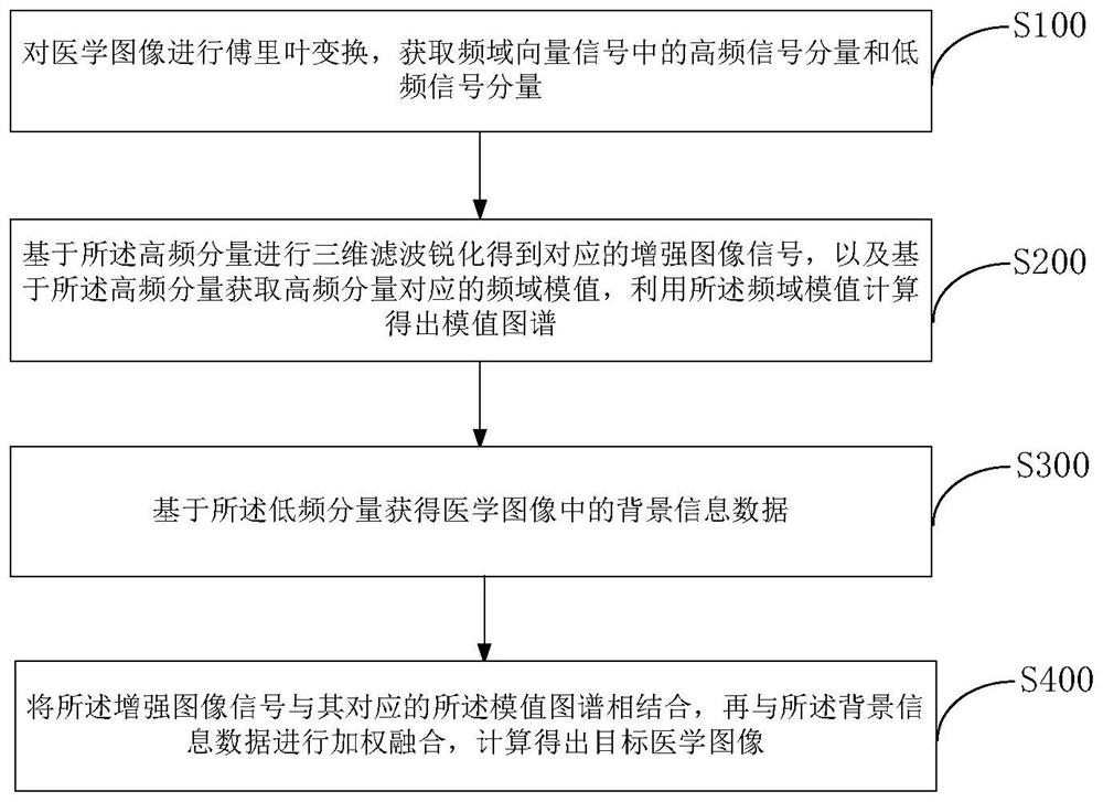
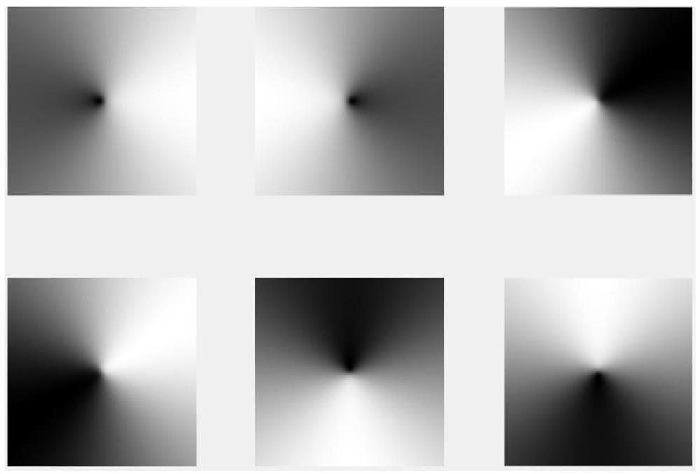
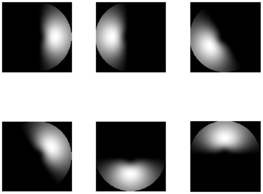
[0043] see figure 1 , figure 1 It is a schematic flowchart of an embodiment of a medical image processing method in an embodiment of the present invention. The medical image processing method extracts the middle and high frequency information from the frequency domain of the medical image, performs three-dimensional filtering and sharpening in six directions to highlight tissue details, and then performs low-frequency filtering on the image to obtain background information, a...


PUM


Abstract

The invention discloses a medical image processing method and device, and the method comprises the steps: carrying out the Fourier transform of a medical image, and obtaining a high-frequency signal component and a low-frequency signal component in a frequency domain vector signal; performing three-dimensional filtering sharpening based on the high-frequency component to obtain a corresponding enhanced image signal, obtaining a frequency domain module value corresponding to the high-frequency component based on the high-frequency component, and calculating by using the frequency domain module value to obtain a module value map; obtaining background information data in the medical image based on the low-frequency component; and combining the enhanced image signal with the corresponding module value map, performing weighted fusion with the background information data, and calculating to obtain a target medical image.

Description

technical field [0001] The invention relates to the field of computer technology, in particular to a method and device for processing medical images. Background technique [0002] When imaging with magnetic resonance technology, the image generated by the original data will be affected by factors such as background noise, artifacts, and boundary blur effects, which will lead to distortion of the tissue structure information of the image, which will not be conducive to the subjective judgment of the doctor. The original image obtained by scanning is processed after filtering and sharpening to reduce the unfavorable image while highlighting the anatomical structure between the image tissues. [0003] Because the original MRI images often have background noise or unfavorable factors of the scanning system (such as inhomogeneity of the magnetic field, output distortion of the gradient coil due to eddy current generated by high-frequency oscillation), the tissue signal is not cle...

Claims


Application Information

Patent Timeline
Patent Type & Authority Applications(China)
IPC IPC(8): G06T5/00G06T5/10G06T5/20A61B5/00A61B5/055
CPCG06T5/10G06T5/20A61B5/055A61B5/0033G06T2207/20056G06T2207/10088G06T2207/20024G06T2207/20221G06T5/73
Inventor 江葵刘省李兰
Owner SHENZHEN BASDA MEDICAL APP
Learn More