Endoscopic image processing method and system, storage medium and equipment

An image processing and endoscopy technology, applied in image data processing, endoscopy, image enhancement, etc., can solve the problems of image reviewers' workload, blurred image quality, occupying database storage capacity, etc., so as to reduce the occupation of database storage disks. Effects of space problems

Pending Publication Date: 2022-05-31
SHANDONG UNIV QILU HOSPITAL
View PDF0 Cites 0 Cited by
  • Summary
  • Abstract
  • Description
  • Claims
  • Application Information

AI Technical Summary

Problems solved by technology

[0003] Endoscopic images are often retained during endoscopic examination. However, as a non-rigid structure, the digestive tract often wriggles, and artifacts, blurring, and other poor image quality often occur when taking pictures.
The inventor found that at present, the method of continuous multi-image acquisition of the same part is often used in clinical practice to solve this problem, and these low-quality images occupy the storage capacity of the database on the one hand, and on the other hand bring a workload to the image review personnel

Method used


Image

  • Endoscopic image processing method and system, storage medium and equipment
  • Endoscopic image processing method and system, storage medium and equipment
  • Endoscopic image processing method and system, storage medium and equipment

Examples


Embodiment 1

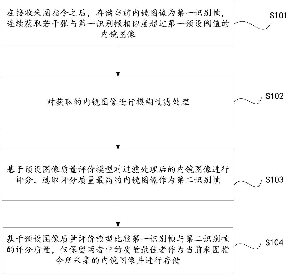
[0040] refer to figure 1 , this embodiment provides an endoscopic image processing method, which specifically includes the following steps:

[0041] S101: After receiving the image acquisition instruction, store the current endoscopic image as the first identification frame, and continuously acquire several endoscopic images whose similarity with the first identification frame exceeds a first preset threshold.

[0042] Wherein, the first preset threshold is preset by humans, and those skilled in the art can perform matching settings according to actual conditions, which will not be described in detail here.

[0043] In this embodiment, the similarity is represented by the Hamming distance.

[0044] Calculate the similarity between two images by calculating the Hamming distance. First, the collected endoscopic images are preprocessed to remove the black border and only keep the valid area. Convert the color image of the effective area to a grayscale image, then scale it to a...

Embodiment 2

[0062] refer to figure 2 , this embodiment is based on the first embodiment, the endoscopic image processing method further includes:

[0063] S105 : After the endoscopy is completed, filter out the repeated endoscopic images whose similarity exceeds the second preset threshold and store the repeated filtered endoscopic images under the same detection site collected by the corresponding image acquisition instruction.

[0064] Wherein, the second preset threshold is preset by humans, and those skilled in the art may perform matching settings according to actual conditions, which will not be described in detail here.

[0065] In the specific implementation process of step S105, when any of the repeatedly filtered endoscopic images obtained has a lesion target, all the repeatedly filtered endoscopic images are retained and stored.

[0066] When there is no focal target in all the repeatedly filtered endoscopic images obtained, from the repeatedly filtered endoscopic images, the...

Embodiment 3

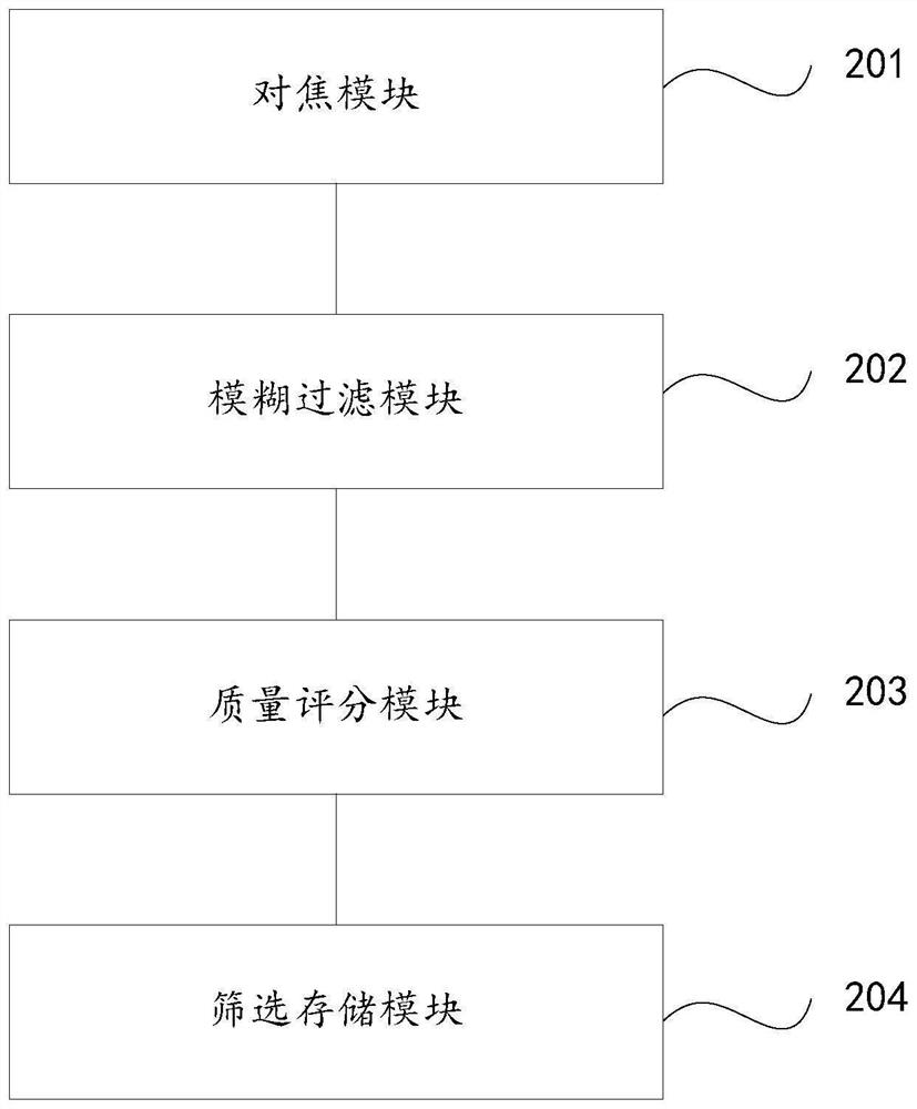
[0072] refer to image 3 , this embodiment provides an endoscopic image processing system, which specifically includes the following modules:

[0073] The focusing module 201 is used to store the current endoscopic image as the first identification frame after receiving the image acquisition instruction, and continuously acquire several endoscopic images whose similarity with the first identification frame exceeds the first preset threshold;

[0074] a blur filtering module 202, which is used to perform blur filtering processing on the acquired endoscopic image;

[0075] A quality scoring module 203, configured to score the filtered endoscopic image based on a preset image quality evaluation model, and select the endoscopic image with the highest scoring quality as the second identification frame;

[0076] Screening storage module 204, which is used to compare the scoring quality of the first recognition frame and the second recognition frame based on a preset image quality e...


PUM


Abstract

The invention belongs to the technical field of endoscope image processing, and provides an endoscope image processing method and system, a storage medium and equipment. In order to solve the problem that low-quality images occupy the storage capacity of a database and bring workload to image auditing personnel, the endoscopic image processing method comprises the steps that after an image acquisition instruction is received, a current endoscopic image is stored as a first recognition frame, and a plurality of endoscopic images with the similarity with the first recognition frame exceeding a first preset threshold value are continuously acquired; performing fuzzy filtering processing on the acquired endoscopic image; scoring the filtered endoscope images based on a preset image quality evaluation model, and selecting the endoscope image with the highest scoring quality as a second recognition frame; and comparing the score quality of the first identification frame and the score quality of the second identification frame based on a preset image quality evaluation model, and only reserving one of the two frames with the best quality as an endoscopic image acquired by the current image acquisition instruction and storing the endoscopic image. The workload of image auditing personnel can be reduced after the number of acquired images is reduced.

Description

technical field [0001] The invention belongs to the technical field of mirror image processing, and in particular relates to an endoscope image processing method, system, storage medium and device. Background technique [0002] The statements in this section merely provide background information related to the present invention and do not necessarily constitute prior art. [0003] Endoscopic images are often retained during endoscopy. However, as a non-rigid structure, the digestive tract often wriggles, resulting in poor image quality such as artifacts and blurring when taking pictures. The inventors found that at present, the method of continuous multi-image acquisition of the same part is often used in clinical practice to solve this problem, and these low-quality images occupy the storage capacity of the database on the one hand, and bring work burden to the image reviewers on the other hand. SUMMARY OF THE INVENTION [0004] In order to solve the technical problems e...

Claims


Application Information

Patent Timeline
Patent Type & Authority Applications(China)
IPC IPC(8): G06T5/00G06N3/08G06N3/04G06K9/62A61B1/273A61B1/00G06V10/764G06V10/74G06V10/82
CPCG06T5/003G06T5/002G06N3/08A61B1/00009A61B1/273G06T2207/30168G06N3/045G06F18/22G06F18/241
Inventor 刘静李真马铭骏赖永航左秀丽李延青陈栋栋姜建科张晨晨
Owner SHANDONG UNIV QILU HOSPITAL
Why Eureka
Try Eureka