Supercharge Your Innovation With Domain-Expert AI Agents!

Knee joint MRI bone structure segmentation method based on 2D-3D feature fusion

A 2D-3D, feature fusion technology, applied in image analysis, neural architecture, image enhancement, etc., can solve the problems of low segmentation accuracy, irregular bone boundaries, narrow gaps at joint joints, etc., to improve practicability and segmentation accuracy. high effect

Pending Publication Date: 2022-05-31
FUDAN UNIV
View PDF0 Cites 0 Cited by
  • Summary
  • Abstract
  • Description
  • Claims
  • Application Information

AI Technical Summary

Problems solved by technology

[0004] The purpose of the present invention is to solve two problems existing in the segmentation of the bone structure of the knee joint based on MR images: 1. The bone structure of the knee joint has extremely irregular bone boundaries, narrow gaps at joint joints, and complex cartilage wear conditions
In view of the low accuracy of 3D-Unet segmentation based on the previous deep learning method, the new method uses the 2D network to extract the global positioning features of the maximum density projection image of the volume data to enrich the global context information missing in the 3D network, so as to improve the bone structure. Segmentation accuracy. In addition, this method of supplementing global information can effectively deal with the problem of a large number of missing semantics caused by the small input block of the 3D network limited by insufficient GPU memory.

Method used


Image

  • Knee joint MRI bone structure segmentation method based on 2D-3D feature fusion
  • Knee joint MRI bone structure segmentation method based on 2D-3D feature fusion
  • Knee joint MRI bone structure segmentation method based on 2D-3D feature fusion

Examples


Embodiment Construction

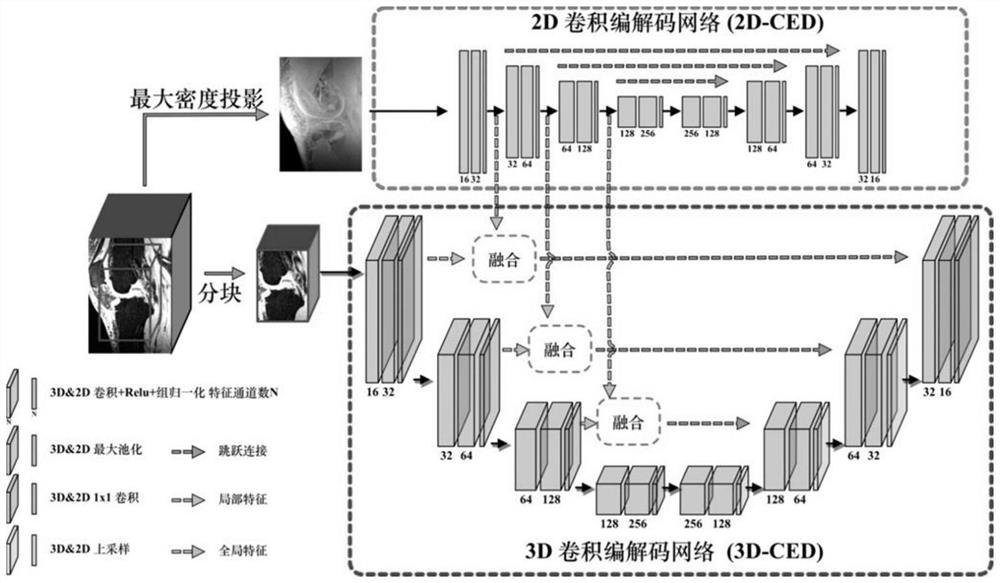
[0030] The overall structure of the method of the present invention is as follows figure 1 As shown, unlike the previous deep learning-based methods, in order to efficiently and accurately segment the bone structure of the knee joint, we propose a new segmentation network architecture based on 2D-3D feature hierarchical fusion.

[0031] 1. Segmentation network based on 2D-3D feature level fusion

[0032] Previous deep learning-based knee bone structure segmentation methods were mainly based on 2D convolutional neural networks, 2D or 3D network hybrid cascades, and neural network segmentation methods combined with anatomical prior information. The new network is a segmentation network based on the features of 2D network and 3D network after hierarchical fusion. The detailed structure is as follows: figure 1As shown, the input of the network is the MR image of the 3D knee joint part, and the output is the segmentation result of the corresponding femur, femoral cartilage, tibia,...


PUM


Abstract

The invention discloses a knee joint MRI bone structure segmentation method based on 2D-3D feature fusion. The method comprises the following steps: firstly, calculating a maximum density projection image (MIP) in a sagittal orientation of MR data, thereby constructing a high-precision convolutional coding and decoding neural network architecture for automatically segmenting knee joints: 1) extracting a 2D bypass network of global features based on the MIP; 2) a 3D backbone network for extracting local detail features based on MR; and 3) a feature fusion module for 2D global information and 3D local detail information. Particularly, the global features serve as position information and are fused with the local detail network at each resolution of a coding path, context information of the local network is increased, and segmentation precision is improved. According to the method, the average dice similarity coefficients of the femur, the femur cartilage, the tibia and the tibia cartilage are as high as 97.78%, 84.83%, 97.93% and 84.80% through verification on a disclosed data set, and the segmentation performance is obviously superior to that of other methods.

Description

technical field [0001] The invention belongs to the technical field of nuclear magnetic resonance imaging (Magnetic Resonance Imaging, MRI) knee joint bone segmentation, and in particular relates to a knee joint MRI bone structure segmentation method based on 2D-3D feature fusion. Background technique [0002] Automatic segmentation of knee bone structures (tibia, tibial cartilage, femur, femoral cartilage) is an important task when diagnosing knee joint diseases based on MRI images. However, the general segmentation method is extremely challenging to accurately segment the joint structure because it cannot take into account both the thick bone and the fine cartilage. At present, the main problems of knee bone segmentation are as follows: 1. In MRI images, there are extremely irregular bone boundaries, narrow gaps at joint joints, and complex cartilage wear in the bone structure of the knee joint. 2. Due to the limitation of the current GPU memory, the three-dimensional (Th...

Claims


Application Information

Patent Timeline
IPC IPC(8): G06T7/00G06T7/10G06V10/80G06V10/82G06K9/62G06N3/04
CPCG06T7/0012G06T7/10G06T2207/10088G06T2207/30008G06T2207/10012G06N3/045G06F18/253
Inventor 史勇红李文生姚德民王辉
Owner FUDAN UNIV
Features
  • R&D
  • Intellectual Property
  • Life Sciences
  • Materials
  • Tech Scout
  • Unparalleled Data Quality
  • Higher Quality Content
  • 60% Fewer Hallucinations
Learn More