
Visual bladder blood clot removing instrument

A technology for removing devices and blood clots, applied in the field of medical devices, can solve problems such as difficulty in grasping the length of the bladder, increasing pressure, and low efficiency

Pending Publication Date: 2022-06-21
徐小娜 +2
View PDF0 Cites 0 Cited by
  • Summary
  • Abstract
  • Description
  • Claims
  • Application Information

AI Technical Summary

Problems solved by technology

When using Ellik, sealing the stuffed bladder will invisibly increase the pressure, causing the bladder to rupture. It is reported that there is a 2.2% risk of rupture. Therefore, after each flushing of blood clots, the gas and liquid in the bladder should be released before the next flushing
[0004] Clinically, blood clots in the bladder are removed by quickly and repeatedly pulling and inserting the obturator with the Ellik flushing ball. During the process of rapidly and repeatedly pulling and inserting the obturator, the irrigation fluid splashes around the surgical area. Endoscopic instruments cut blood clots into small strips and use Ellik flushing balls to suck them out, but the efficiency is very low
Yu Limin and others used endoscopic aspirator to remove bladder blood clots, but the endoscopic aspirator is a metal straight rod, the length of penetration into the bladder is difficult to grasp, easy to puncture the bladder, and the safety is low; Hong Yude et al. used Cook ureteral delivery sheath to remove bladder blood clots, but Cook ureteral delivery sheath is a high-tech consumable, which is expensive and increases the financial burden of patients; Apul Goel et al. studied a new device with a vent hole on the top between the outer sheath of the resectoscope and the suction tube. Intermittent mechanical suction, which can maintain the pressure balance in the bladder; most of the methods are to achieve the purpose of sucking blood clots by changing the suction device to cooperate with the negative pressure suction device, but they are all operated blindly

Method used


Image

  • Visual bladder blood clot removing instrument
  • Visual bladder blood clot removing instrument
  • Visual bladder blood clot removing instrument

Examples


Embodiment Construction

[0023] The technical solutions in the embodiments of the present invention will be clearly and completely described below with reference to the accompanying drawings in the embodiments of the present invention. Obviously, the described embodiments are only a part of the embodiments of the present invention, but not all of the embodiments. Based on the embodiments of the present invention, all other embodiments obtained by those of ordinary skill in the art without creative efforts shall fall within the protection scope of the present invention.

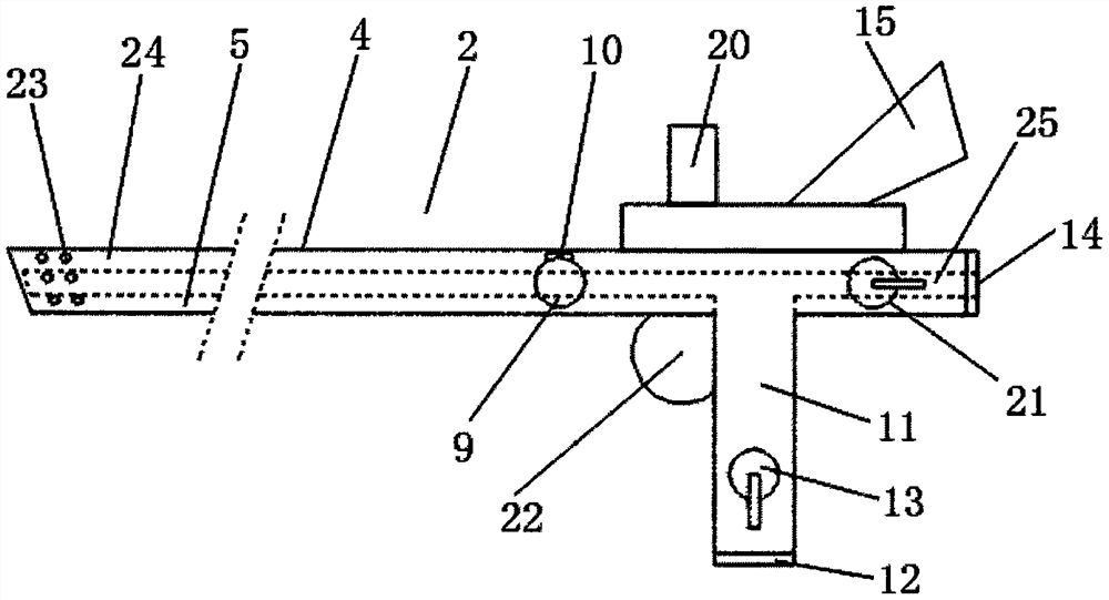
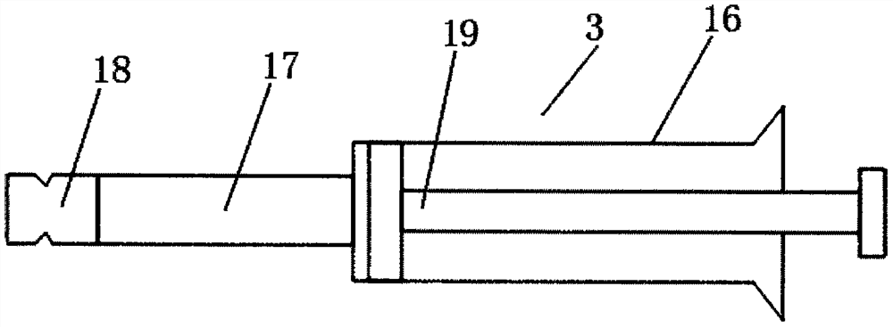
[0024] see Figure 1-4 , a visual bladder blood clot removal device, including an obturator 1, a lens 2, a suction syringe 3, the lens 2 includes a mirror rod 4, the outer wall of the mirror rod 4 is a smooth curved surface, and the interior of the mirror rod 4 is provided with a flushing work Channel 5, a light guide 24 is provided above the flushing working channel 5, a flushing outlet 6 is provided on the left side of the flushing ...


PUM


Abstract

The invention relates to the field of medical instruments, in particular to a visible bladder blood clot removing instrument which comprises an obturator, a lens and a suction injector, a flushing outlet is formed in the left side of a flushing working channel, an objective lens is arranged above the flushing outlet, a suction connector is formed in the right side of the flushing working channel, and a camera connector is installed above a lens rod. The instrument has the advantages that the instrument has a visual function through the objective lens, the camera interface and the cold light source interface, the instrument has two modes and functions, a suction mode or a suction mode can be selected according to blood clot properties, and the suction mode is suitable for sparse blood clots with short blood clot forming time; the suction mode is suitable for large-area coagulated compact organic blood clots which are long in blood clot forming time, so that large equipment and large instruments are not needed in the operation process, the burden of itinerant nurses is not increased, fixing equipment and instruments in a ready-made operating room can be utilized, and the convenient, safe and efficient performance is achieved.

Description

technical field [0001] The invention relates to the field of medical devices, in particular to a visible bladder blood clot removal device. Background technique [0002] Bladder clot packing is a very common emergency in urology. The common causes are urinary system trauma such as renal contusion, bladder contusion, postoperative bleeding such as transurethral resection of bladder tumor and resection of prostate, and bladder irritation. inflammation, after intravesical chemotherapy. The onset of bladder clot tamponade is acute, and the patient presents with dysuria, severe abdominal pain, obvious abdominal distension, and extreme pain. Conservative treatment is often required, such as inserting a three-lumen catheter for continuous bladder irrigation. If conservative treatment fails, emergency surgery is required. These blood clot tamponade patients who need surgical treatment often have a long onset time, large blood clot volume, and a large number of organized blood clots...

Claims


Application Information

Patent Timeline
IPC IPC(8): A61M1/00A61B1/307
CPCA61B1/307A61M2205/583A61M2210/1085A61M2210/005
Inventor 徐小娜徐自强黄崇础
Owner 徐小娜
Learn More