
Method and apparatus for segmenting structure in CT angiography

一种CT图像、脉管的技术,应用在体积医学成像,医学成像领域,能够解决不能获得体积数据组优点、不管数据可用性、妨碍其它模糊的病理发现等问题

Inactive Publication Date: 2005-06-01
GENERAL ELECTRIC CO
View PDF0 Cites 41 Cited by
  • Summary
  • Abstract
  • Description
  • Claims
  • Application Information

AI Technical Summary

Problems solved by technology

As a result, the operator may have to repeatedly re-draw or re-contour the entire procedure, possibly taking more than 1 hour to process the imaged head and neck volume
Furthermore, operator intervention may lead to variability in the segmentation of structures between users and within the same user
[0008] Since this procedure is time consuming, the operator may attempt to reduce processing time by restricting processing to regions or volumes of interest corresponding to the location of the pathology
However, limiting the processing in this way does not take full advantage of all available volume data sets
Furthermore, restricting processing to regions of known or suspected pathology may have hampered the discovery of otherwise obscure pathologies, regardless of data availability

Method used


Image

  • Method and apparatus for segmenting structure in CT angiography
  • Method and apparatus for segmenting structure in CT angiography
  • Method and apparatus for segmenting structure in CT angiography

Examples


Embodiment Construction

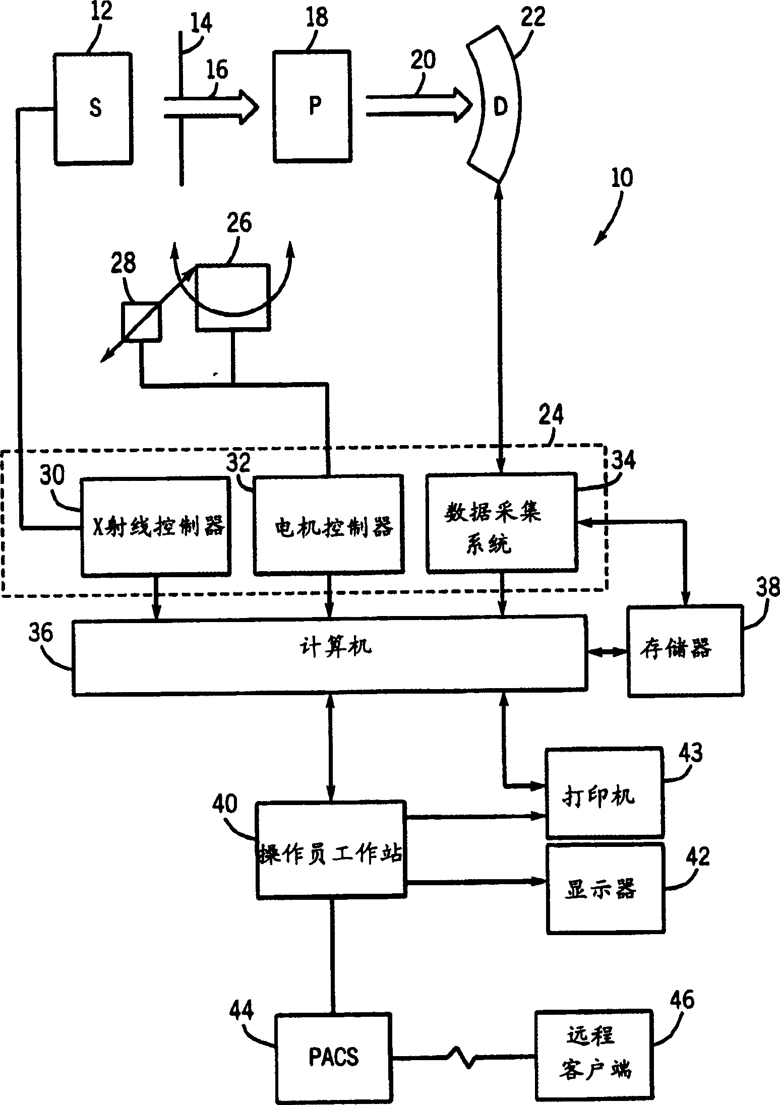
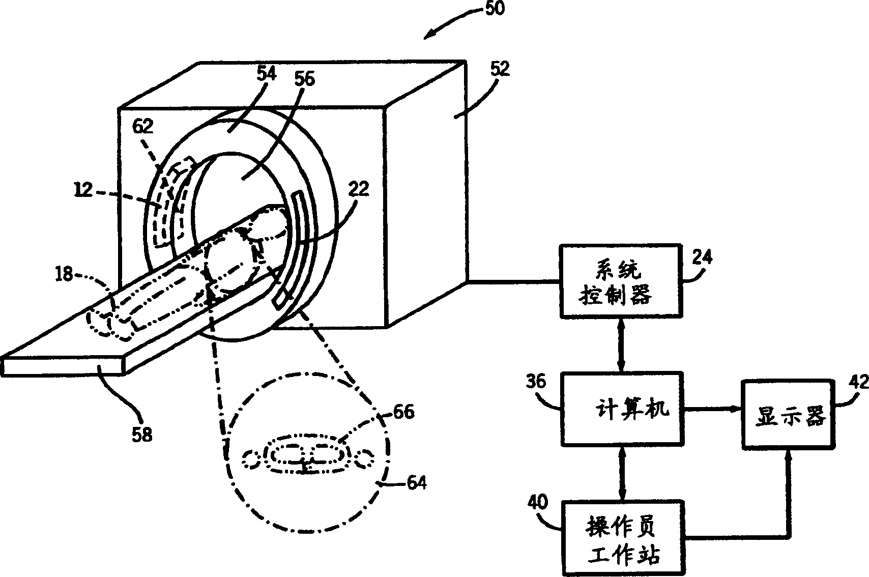
[0016] figure 1 The imaging system 10 for acquiring and processing image data is illustrated diagrammatically. In the illustrated embodiment, the system 10 is a computer tomography (CT) system according to the present technology, which is designed to not only collect raw image data but also process the image data for display and analysis. Other imaging modes for acquiring volumetric image data, such as magnetic resonance imaging (MRI) or positron emission tomography (PET), can also benefit from this technology. The following discussion on the CT system is only an example of such an implementation, and is not limited by mode or anatomical location.

[0017] in figure 1 In the illustrated embodiment, the imaging system 10 includes an X-ray radiation source 12 positioned adjacent to the collimator 14. In this exemplary embodiment, the source of X-ray radiation source 12 is typically an X-ray tube. The collimator 14 allows the radiation beam 16 to penetrate the area where the subje...


PUM


Abstract

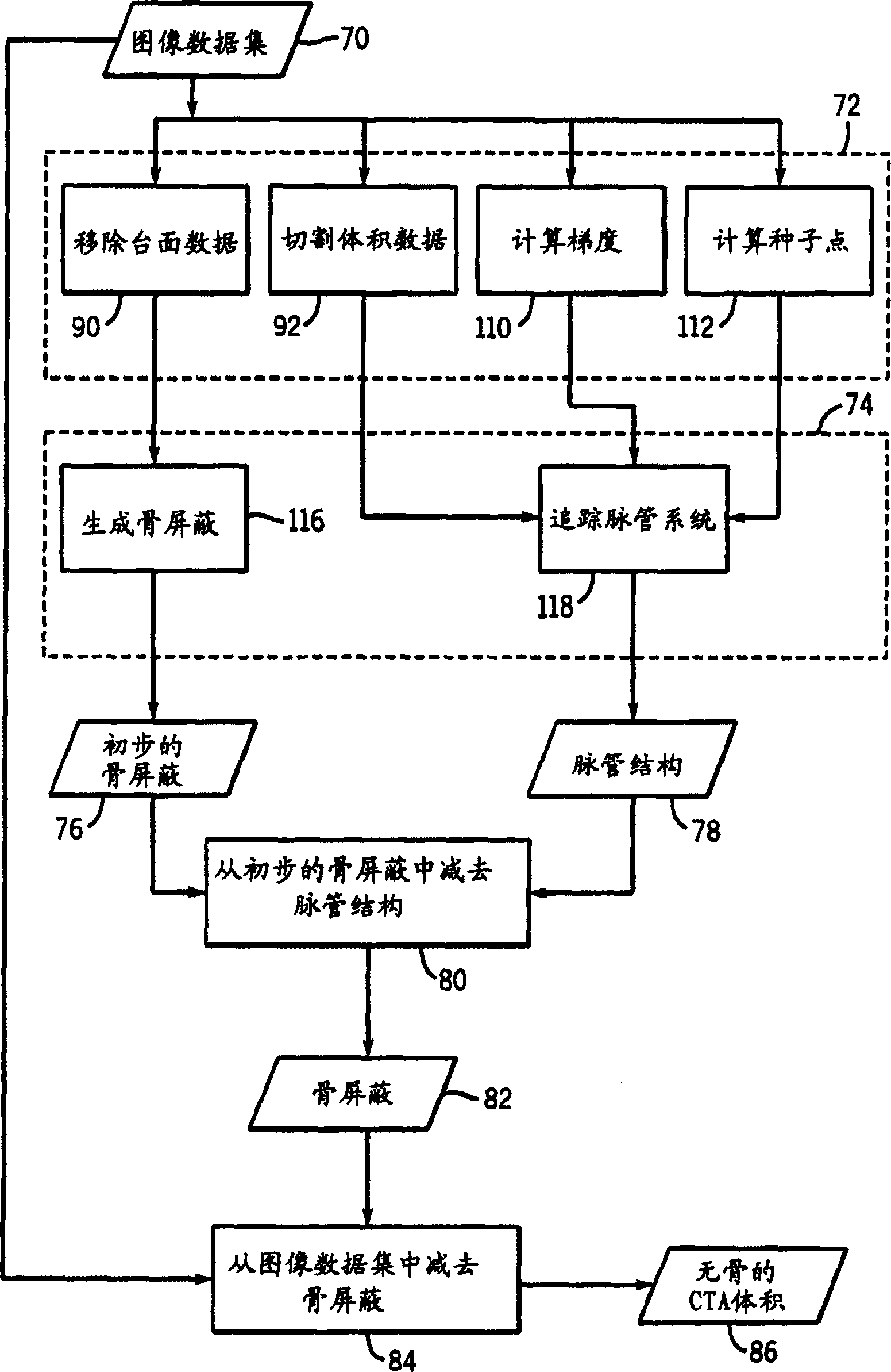
The present invention provides a technique for automatically generating bone shielding (82) in CTA angiography. This technique can preprocess the image data set (70) to perform various functions, such as removing (90) the image data associated with the table top, cutting (92) the volume into regionally consistent sub-volumes, calculating according to the gradient ( 110) Structure edges, and / or computing (112) seed points for subsequent region growing. The preprocessed data can then be automatically segmented for bone and vascular structures (78). This automatic vessel segmentation (118) can be accomplished using suppression region growing, where the suppression is dynamically updated according to the local statistics of the image data. The vasculature (78) may be subtracted (80) from the bone structure (76) to generate a bone shield (82). The bone mask (82) may in turn be subtracted (84) from the image dataset (70) to generate a boneless CTA volume (86) for volume-rendered reconstruction.

Description

Technical field [0001] The present invention generally relates to the field of medical imaging, and particularly relates to the field of volumetric medical imaging. In particular, the present invention relates to a technique for segmenting bone and vasculature data in computed tomography. Background technique [0002] Volumetric medical imaging technology uses a variety of technologies to gather three-dimensional information of the human body. For example, computed tomography (CT) imaging systems measure the attenuation of X-ray beams passing through a patient from multiple angles. Based on these measurements, the computer can reconstruct the image of the patient's body that caused the radiation attenuation. As those skilled in the art will understand, these images are individually detected based on a series of angularly displaced cross sections. It should be pointed out that the CT system generates data representing the distribution of the linear attenuation coefficient of the s...

Claims


Application Information

Patent Timeline
Patent Type & Authority Applications(China)
IPC IPC(8): A61B6/03A61B6/00G06T5/00
CPCA61B6/032A61B6/4085A61B6/481A61B6/504G06T5/50G06T7/0012G06T2207/10081G06T2207/20012G06T2207/20156G06T2207/20224G06T2207/30008G06T2207/30101G06T7/11G06T7/12G06T7/174G06T7/187G06V40/14G06V10/273G06V2201/03
Inventor S·叙尔亚纳拉亚南R·穆利克Y·马尔亚V·P·卡马特N·纳加拉
Owner GENERAL ELECTRIC CO
Learn More