Methods and devices for draining fluids and lowering intraocular pressure

A shunt device and liquid technology, applied in the fields of medicine and surgery, can solve the technical difficulties of filter tract angioplasty, high rate of early hypotonic endophthalmitis, etc.

Active Publication Date: 2006-03-22
汉帕尔·卡拉乔济安 +1
View PDF3 Cites 13 Cited by
  • Summary
  • Abstract
  • Description
  • Claims
  • Application Information

AI Technical Summary

Problems solved by technology

However, tractoplasty is technically difficult, fraught with high rates of early hypotonia, late failure, and endophthalmitis leading to permanent blindness of the eye

Method used


Image

  • Methods and devices for draining fluids and lowering intraocular pressure
  • Methods and devices for draining fluids and lowering intraocular pressure
  • Methods and devices for draining fluids and lowering intraocular pressure

Examples


Embodiment Construction

[0027]The following detailed description and the accompanying drawings are provided to illustrate non-limiting embodiments of the present invention only. This detailed description is not intended to describe all examples and implementations of the invention, and therefore, is not intended to limit the invention in any way.

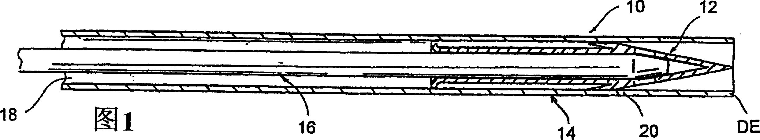
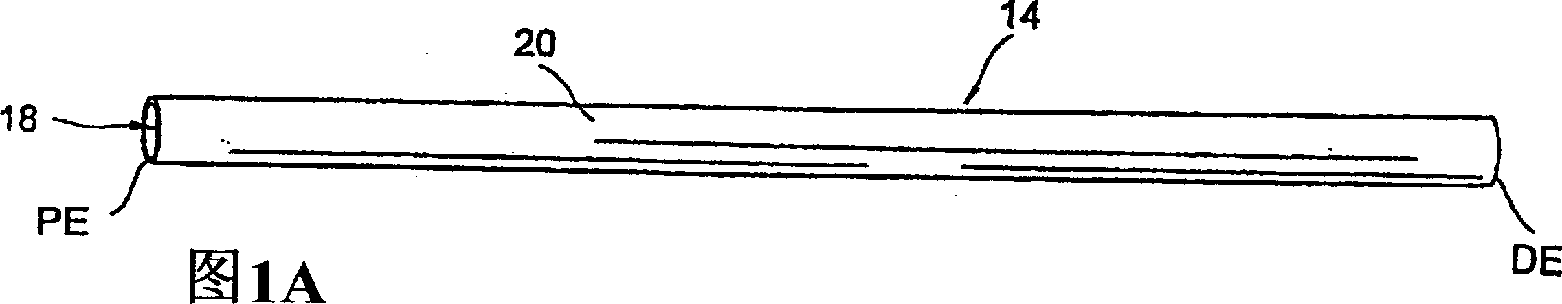
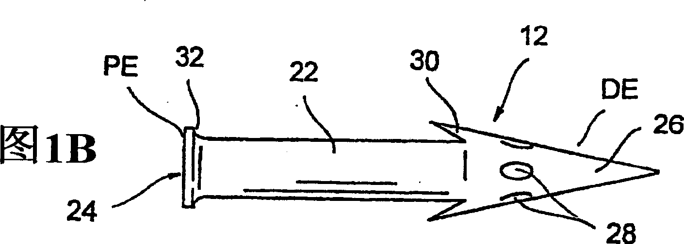
[0028] 1-1C illustrate one embodiment of a system 10 for implanting a shunt device 12 of the present invention. The system 10 generally includes a diverter device 12 , a cannula 14 , and a pusher 16 . As shown in Figure 1, the shunt device 12 is initially located in the lumen 18 of the sleeve 14, and the pusher 16 is positioned behind the shunt device 12 in the lumen 18 of the sleeve 114, so that the shunt device 12 can be moved from the sleeve 14 using the pusher 16. launched in the remote DE. One way to carry out this diversion-drainage process is as Figure 2A shown in and detailed below.

[0029] A particular embodiment of a flow splitting device 1...


PUM


Abstract

Methods, devices and systems for draining fluid from the eye and / or for reducing intraocular pressure. A passageway (e.g., an opening, puncture or incision) is formed in the lamina cribosa or elsewhere to facilitate flow offluid from the posterior chamber of the eye to either a) a subdural location within the optic nerve or b) a location within the subarachnoid space adjacent to the optic nerve. Fluid from the posterior chamber then drains into the optic nerve or directly into the subarachnoid space, where it becomes mixed with cerebrospinal fluid. In some cases, a tubular member (e.g., a shunt or stent) may be implanted in the passageway. A particular shunt device and shunt-introducer system is provided for such purpose. A vitrectomy or vitreous liquefaction procedure may be performed to remove some or all of the vitreous body, thereby facilitating creation of the passageway and / or placement of the tubular member as well as establishing a route for subsequent drainage of aqueous humor from the anterior chamber, though the posterior chamber and outwardly though the passageway where it becomes mixed with cerebrospinal fluid.

Description

[0001] related application [0002] This application claims priority to US Provisional Patent Application Serial No. 60 / 447,999, filed February 18, 2003, which is incorporated herein by reference. technical field [0003] The present invention relates generally to the fields of medicine and surgery, and more particularly to methods and devices for reducing intraocular pressure in human and veterinary patients. Background technique [0004] In a normal adult, the eyeball is approximately spherical, with an average diameter of 24.5 mm. The cornea is a transparent tissue that inserts into the sclera at the limbus, with the anterior chamber behind the cornea. The iris is the anterior extension of the ciliary body, which appears as a plane with a circular opening, the pupil, in the center of this plane. The iris is adjacent to the anterior surface of the lens and separates the anterior chamber from the posterior chamber, which each contain aqueous humor. The lens is a biconvex...

Claims


Application Information

Patent Timeline
Patent Type & Authority Applications(China)
IPC IPC(8): A61M5/00A61F2/14A61B19/00A61FA61F9/007
CPCA61F9/00781
Inventor 汉帕尔·卡拉乔济安雨果·基罗斯-梅尔卡多
Owner 汉帕尔·卡拉乔济安
Why Eureka
Try Eureka