

Method and device for visually assisting the electrophysiological use of a catheter in the heart
A technique for electrophysiology and catheterization, used in applications, measuring devices, utilizing re-radiation, etc.
- Summary
- Abstract
- Description
- Claims
- Application Information
AI Technical Summary
Problems solved by technology
Method used
Image

Examples
Embodiment Construction
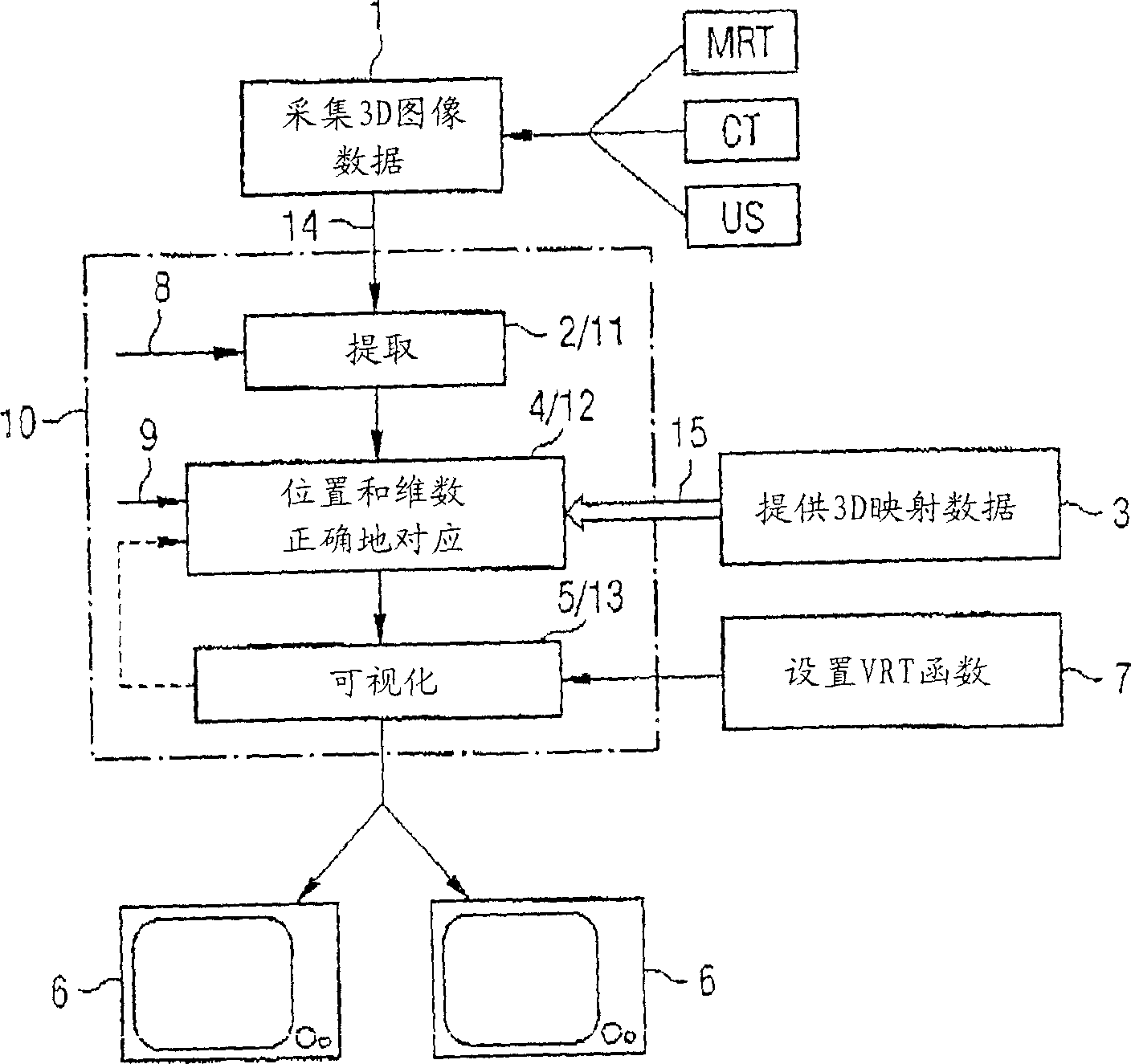
[0020] In the method according to the invention, in a first step 1 3D image data of the body part, in particular containing the ventricle to be treated, are acquired. During the acquisition of the 3D image data, it is also possible for a larger part of the heart to be involved for the recording to be carried out later. The acquisition of the 3D image data is carried out using tomographic 3D imaging methods, such as X-ray computed tomography, magnetic resonance tomography or 3D ultrasound technology. When acquiring the 3D image data, it should be noted that these are acquired in each case for the same cardiac phase for which the electroanatomical 3D mapping data are also provided later. This is ensured by EKG gating of image acquisition as well as EKG gating of 3D mapping data acquisition, eg by reference to a percentage of the RR interval or a fixed time interval before or after the reference R spike.
[0021] When carrying out the method according to the invention it is impo...
PUM
 Abstract
Description
Claims
Application Information