
Apparatus for automatically detecting salient features in medical image data

An automatic detection, medical image technology, applied in image data processing, image data processing, image enhancement and other directions, can solve problems such as neglect

Active Publication Date: 2007-03-28
SIEMENS HEALTHCARE GMBH
View PDF0 Cites 9 Cited by
  • Summary
  • Abstract
  • Description
  • Claims
  • Application Information

AI Technical Summary

Problems solved by technology

Although diagnostically significant abnormalities in the recorded image data can be identified by an experienced user, there is a danger in the case of an inexperienced user that these abnormalities are caused by a not always optimal image quality. being ignored

Method used


Image

  • Apparatus for automatically detecting salient features in medical image data

Examples


Embodiment Construction

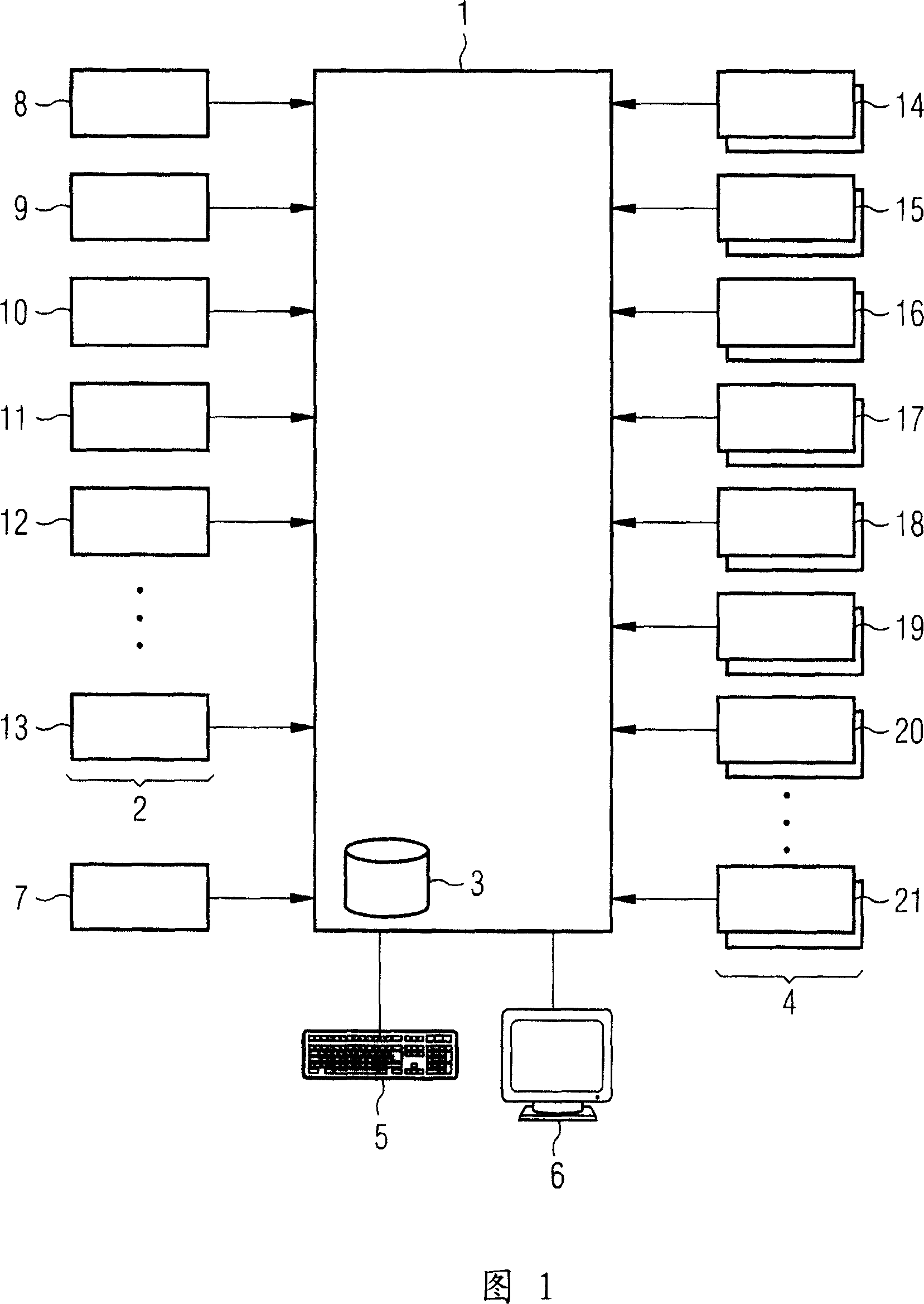
[0013] 1 here shows an embodiment of such a device with a control unit 1, one or more determination modules 2, a memory unit 3 for storing image data, a plurality of examination modules 4, an input unit 5 for the user and as Monitor 6 of the output unit. It can be seen in this embodiment that one inspection module 14 in the inspection modules 4 has one or more application programs for detecting human organ damage, one inspection module 15 has one or more application programs for detecting embolism, An inspection module 16 has one or more application programs for detecting stenosis, an inspection module 17 has one or more application programs for detecting lung soft tissue diseases, and an inspection module 18 has one or more application programs for detecting bone quality An examination module 19 has one or more applications for detecting aneurysms and an examination module 20 has one or more applications for detecting anatomical defects. Further examination modules with othe...


PUM


Abstract

An apparatus is disclosed for automatically detecting salient features in medical image data. The apparatus includes a memory device for storing the image data, at least one determination module for determining one or more anatomical regions which are acquired by the image data, a number of different examination modules that respectively include an application for automatically detecting specific salient features in a specific anatomical region, an input unit via which a primary application can be started, a control unit that on the basis of the anatomical regions determined by the determination module automatically selects and executes in the background further applications, as well as an output unit on which the result of the primary application is displayed together with an item of information relating to additional salient features that have been detected automatically with the aid of the applications executed in the background. The apparatus improves the diagnostic evaluation of medical image data.

Description

technical field [0001] The invention relates to a device for automatically detecting anomalies in medical image data of a body part of a patient, having storage means for storing the image data, input means capable of launching an application for automatically detecting specific anomalies in the image data, and An output device that displays the results of this application. Background technique [0002] Medical imaging is used in a wide variety of diagnostic problems to support the diagnosis of patients. Although diagnostically significant abnormalities in the recorded image data can be identified by an experienced user, there is a danger in the case of an inexperienced user that these abnormalities are caused by a not always optimal image quality. and was ignored. In order to reduce the occurrence of such problems, devices that can automatically detect abnormalities in recorded image data are known. These devices generally comprise a data processing station with an image...

Claims


Application Information

Patent Timeline
Patent Type & Authority Applications(China)
IPC IPC(8): A61B6/03G06F19/00G06T1/00G06T7/60
CPCG06T2207/30004G06T7/0012A61B6/032
Inventor 卢茨·冈德尔丹尼尔·林克
Owner SIEMENS HEALTHCARE GMBH
Learn More