Analysis of sea-horse quantitative shape in magnetic resonance image

A technology of magnetic resonance data and hippocampus, which is applied in diagnostic recording/measurement, medical science, sensors, etc., can solve problems such as inaccuracy, poor effect, and influence on the credibility of statistical analysis results

Inactive Publication Date: 2007-04-04
INST OF AUTOMATION CHINESE ACAD OF SCI
View PDF0 Cites 6 Cited by
  • Summary
  • Abstract
  • Description
  • Claims
  • Application Information

AI Technical Summary

Problems solved by technology

The registration method based on the overall shape of the hippocampus is usually based on grayscale and feature-based, so the effect of applying it to hippocampus registration is not good, which directly affects t

Method used


Image

  • Analysis of sea-horse quantitative shape in magnetic resonance image
  • Analysis of sea-horse quantitative shape in magnetic resonance image
  • Analysis of sea-horse quantitative shape in magnetic resonance image

Examples


Embodiment Construction

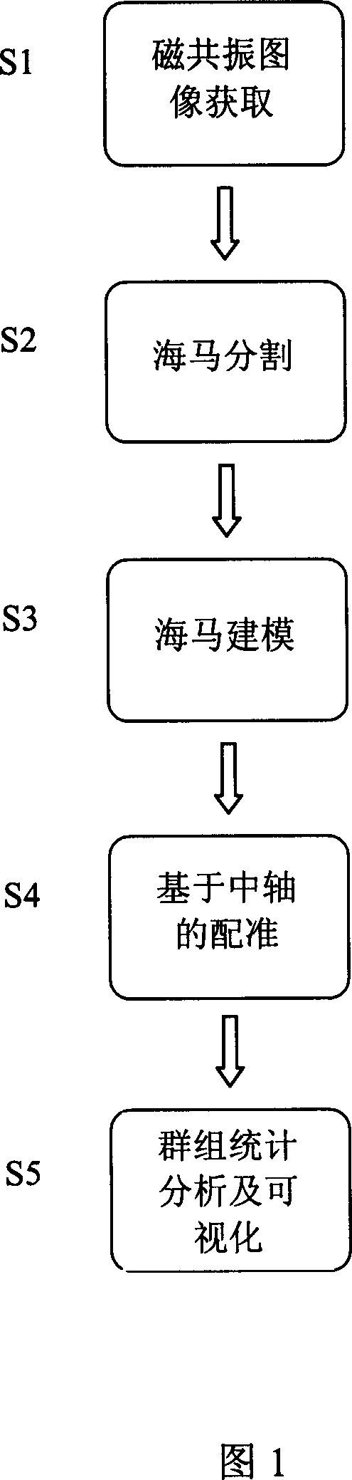
[0021] As shown in Figure 1:

[0022] Step 1 (S1): Magnetic resonance image acquisition.

[0023] The usual high-resolution 3-dimensional T1-weighted images are scanned by spin echo sequence (SE) or gradient echo sequence (SPGR) at 1.5 Tesla, and the spatial resolution is on the order of millimeters. With the development of magnetic resonance imaging technology, there are more and more scans using 3Tesla magnetic field strength. This scan method has higher resolution and clearer images.

[0024] Step 2 (S2): hippocampus segmentation.

[0025] The hippocampus itself belongs to the gray matter. It is almost the same gray scale as the surrounding tissue, and it is difficult to distinguish. The amygdala, another gray matter structure in the head, is even connected to each other, which brings great difficulties to the segmentation of the hippocampus. At present, the automatic segmentation method based on grayscale, feature and prior knowledge is not mature enough, and the accurac...


PUM


Abstract

A method for quantitatively analyzing the hippocampal shape in magnetic resonance image includes such steps as acquiring the data of magnetic resonance image, pre-processing, manually dividing the hippocampus, extracting the coordinates of the dots on the hippocampal surface by marching cube method, creating a parameterized curved surface model of hippocampus, finding out the central axis of hippocampus, using the area of relative parallel coil for registration to the central axis to obtain better tissue correspondency, and significant statistical analyzing by arranging-testing method.

Description

technical field [0001] The invention relates to the field of magnetic resonance image post-processing, and relates to a group shape analysis method based on the hippocampal axis in the magnetic resonance image, which processes the magnetic resonance data of the brain. Background technique [0002] There are two hippocampus-shaped gray matter structures in the human brain. They are located at the back of the brain, one on the left and one on the left. Anatomically called the hippocampus, they are closely related to human cognition and memory functions. Early research on the hippocampus was limited to autopsy, and the method was relatively simple and difficult to apply to the early diagnosis and clinical treatment of the hippocampus. Since the 1980s, with the development of medical imaging technology, magnetic resonance imaging technology has been applied clinically. Magnetic resonance imaging (Magnetic resonance imaging, MRI) is an imaging technique that uses the signal gene...

Claims


Application Information

Patent Timeline
IPC IPC(8): A61B5/055
Inventor 蒋田仔石峰李淑宇朱礼涛
Owner INST OF AUTOMATION CHINESE ACAD OF SCI
Why Eureka
Try Eureka